

A new simplified sonographic approach for pararadicular injections in the lumbar spine: a CT-controlled cadaver study
- PMID: 21349957
- PMCID: PMC7965556
- DOI: 10.3174/ajnr.A2389
A new simplified sonographic approach for pararadicular injections in the lumbar spine: a CT-controlled cadaver study
Abstract
Background and purpose: Injection therapies play a major role in the treatment of lower back pain and are to date performed mainly under CT or fluoroscopic guidance. The benefits of US-guided instillation procedures have been shown in many studies. We conducted this study to simplify an US-guided approach to the lumbar spinal nerves and to assess the feasibility and preliminary accuracy by means of CT and anatomic dissection.
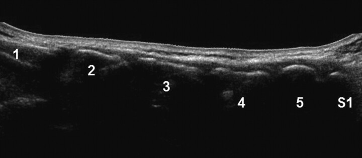
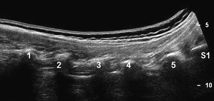
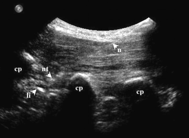
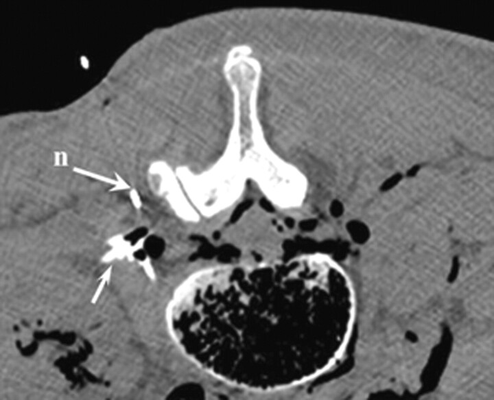
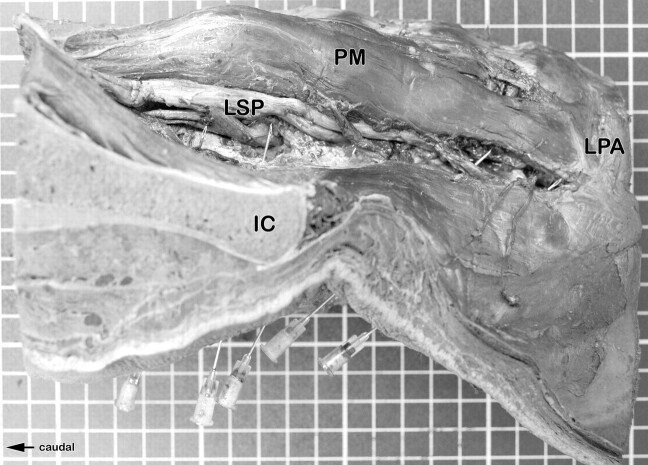
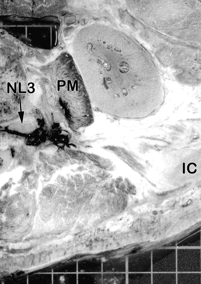
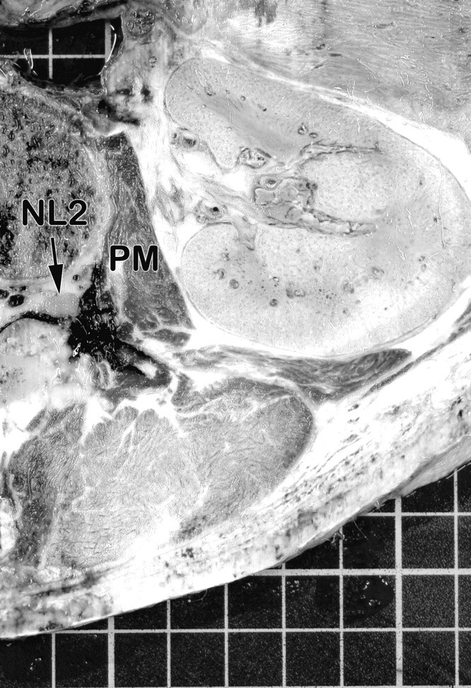
Materials and methods: Ten US-guided injections at 5 different levels (L1-L5) were performed on 1 embalmed cadaver. Images in 3 sagittal/parasagittal scanning planes were obtained at each lumbar level: 1) the plane of the spinous processes, 2) the plane of the lumbar arches/zygapophyseal-joints, and 3) the plane of the transverse processes. The PAP was then defined by positioning the transducer perpendicularly over the medial part of the respective transverse processes, depicting the hyperechoic intertransverse ligament. In the "in-plane technique," spinal needles were advanced through the respective segmental intertransverse ligament. A solution consisting of a contrast agent and a pigmented dispersion was subsequently injected into the pararadicular compartment. An anatomic dissection of the specimen and CT scans were performed to verify the exact placement of the needle tips and to evaluate fluid dispersion in the punctured compartment.
Results: CT examination confirmed that each needle tip was correctly placed within the intended compartment with sufficient contrast accumulation around the respective proximal segment of the spinal nerve. On each anatomic section, dye was identified in the correct compartment and directly around each targeted spinal nerve with needles shown in the correct position.
Conclusions: This modified US approach for therapeutic root injections in the lumbar spine by using the intertransverse ligament as a new anatomic landmark allows an easy and correct needle placement within the pararadicular compartment.
Figures

References
-
- Bogduk N. On the definitions and physiology of back pain, referred pain, and radicular pain. Pain 2009; 147: 17–19 - PubMed
-
- Moore RA, Straube S, Derry S, et al. Chronic low back pain analgesic studies: a methodological minefield. Pain 2010; 149: 431–34 - PubMed
-
- O'Neill S, Graven-Nielsen T, Manniche C, et al. Ultrasound guided, painful electrical stimulation of lumbar facet joint structures: an experimental model of acute low back pain. Pain 2009; 144: 76–83 - PubMed
-
- Schiltenwolf M, Schneider S. Activity and low back pain: a dubious correlation. Pain 200;143:1–2 - PubMed
-
- Carrino JA, Morrison WB, Parker L, et al. Spinal injection procedures: volume, provider distribution, and reimbursement in the US Medicare population from 1993 to 1999. Radiology 2002; 225: 723–29 - PubMed
MeSH terms
LinkOut - more resources
Full Text Sources
Research Materials