

Apparent diffusion coefficients of metabolites in patients with MELAS using diffusion-weighted MR spectroscopy
- PMID: 21349966
- PMCID: PMC7965562
- DOI: 10.3174/ajnr.A2395
Apparent diffusion coefficients of metabolites in patients with MELAS using diffusion-weighted MR spectroscopy
Abstract
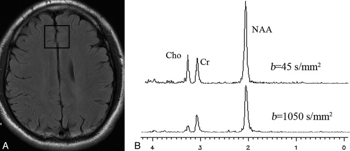
Background and purpose: DW-MR spectroscopy can detect the diffusion coefficients of NAA, Cr, PCr, and Cho and can, therefore, provide some useful information. The aims of this study were to probe the mechanisms underlying the pathogenesis of MELAS and to see whether DW-MR spectroscopy is a useful technique for other diseases besides cerebral infarction.
Materials and methods: Fifteen healthy volunteers and 10 patients with MELAS were enrolled in the study. All were scanned on a 3T whole-body MR imaging scanner. Fifteen ADCs of the singlet metabolites in the gray matter of the healthy subjects, 10 ADCs of the singlet metabolites in the lesions, and 8 ADCs of the singlet metabolites in the nonaffected areas were used in the statistical analysis, respectively.
Results: The metabolite ADCs of the nonaffected areas and the lesions in the patients were higher than those of the frontal gray matter in the healthy controls. There were significant differences between the metabolite ADCs of the nonaffected areas in patients and those in the healthy controls, and it was the same with the metabolite ADCs of the lesions and those of the healthy controls.
Conclusions: The increased ADC values of the metabolites reveal that MELAS is a mitochondrial neuronopathy and involves the entire brain. DW-MR spectroscopy is a very useful noninvasive technique, which can show some valuable information that conventional MR imaging cannot display. Thus, it can be applied to brain diseases besides cerebral infarction.
Figures


References
-
- Stejskal EO, Tanner JE. Spin diffusion measurements: spin echoes in the presence of a time dependent field gradient. J Chem Phys 1965;42:288–92
-
- Busza AL, Allen KL, King MD, et al. Diffusion-weighted imaging studies of cerebral ischemia in gerbils: potential relevance to energy failure. Stroke 1992;23:1602–12 - PubMed
-
- Schaefer PW, Grant PE, Gonzalez RG. Diffusion-weighted MR imaging of the brain. Radiology 2000;217:331–45 - PubMed
-
- Liu Z, Liu X, Hui L, et al. The appearance of ADCs in the non-affected areas of the patients with MELAS. Neuroradiology 2011;53:227–32 - PubMed
-
- Neumann-Haefelin T, Wittsack HJ, Wenserski F, et al. Diffusion- and perfusion-weighted MRI: the DWI/PWI mismatch region in acute stroke. Stroke 1999;30:1591–97 - PubMed
Publication types
MeSH terms
Substances
LinkOut - more resources
Full Text Sources