Case Reports
. 2011 Apr 1;332(6025):65-8.
doi: 10.1126/science.1200439. Epub 2011 Feb 24.

Chronic mucocutaneous candidiasis in humans with inborn errors of interleukin-17 immunity

Affiliations
Case Reports

Chronic mucocutaneous candidiasis in humans with inborn errors of interleukin-17 immunity

Anne Puel et al. Science. .

Abstract

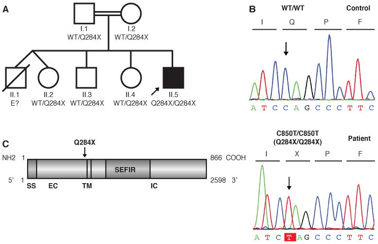
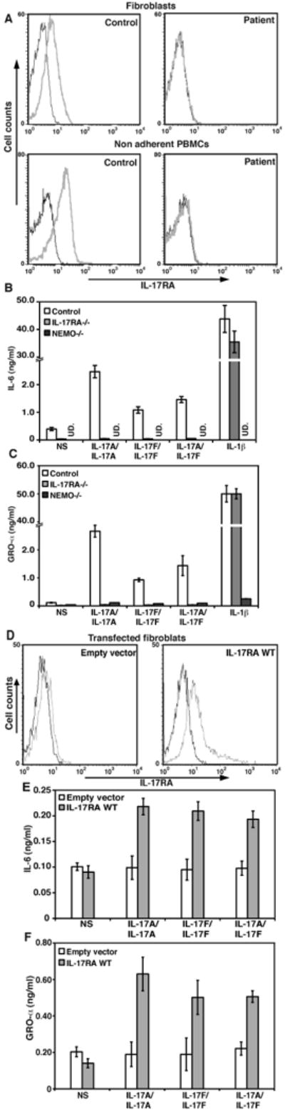
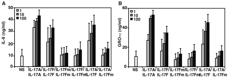
Chronic mucocutaneous candidiasis disease (CMCD) is characterized by recurrent or persistent infections of the skin, nails, and oral and genital mucosae caused by Candida albicans and, to a lesser extent, Staphylococcus aureus, in patients with no other infectious or autoimmune manifestations. We report two genetic etiologies of CMCD: autosomal recessive deficiency in the cytokine receptor, interleukin-17 receptor A (IL-17RA), and autosomal dominant deficiency of the cytokine interleukin-17F (IL-17F). IL-17RA deficiency is complete, abolishing cellular responses to IL-17A and IL-17F homo- and heterodimers. By contrast, IL-17F deficiency is partial, with mutant IL-17F-containing homo- and heterodimers displaying impaired, but not abolished, activity. These experiments of nature indicate that human IL-17A and IL-17F are essential for mucocutaneous immunity against C. albicans, but otherwise largely redundant.


Figures

Fig. 1
Fig. 1
A kindred with autosomal recessive IL-17RA deficiency. (A) Pedigree of the family established by IL-17RA genotyping. The proband is indicated by an arrow. E? indicates individuals whose genetic status could not be evaluated. (B) IL17RA DNA sequence electrophoregrams for a control and the patient. (C) Schematic diagram of the IL-17RA protein with the signal sequence (SS), extracellular (EC), transmembrane (TM), intracellular (IC) and SEFIR (similar expression to FGF/IL-17R) domains, and the position within the extracellular domain affected by the mutation.
Fig. 2
Fig. 2
Production and function of the mutant IL-17RA chain. (A) IL-17RA expression in SV40-immortalized fibroblasts from a control and the patient (top panel) and non adherent PBMCs (lower panel), as detected by flow cytometry. Isotype control (black); IL-17RA antibody (gray). (B) IL-6 and (C) GRO-α production by SV40-immortalized fibroblasts from a control, the patient, and NEMO-deficient cells after 24 hours of stimulation with IL-17A, IL-17F and IL-17A/IL-17F, mean +/- standard deviation (SD) error bars of three independent experiments, as detected by ELISA (UD. Undetectable). (D) IL-17RA expression in SV40-immortalized fibroblasts from the patient, transfected with the empty pORF9mcs plasmid (left panel) or the pORF9-hIL-17RA plasmid (right panel), as detected by flow cytometry. Isotype control (black); IL17RA antibody (gray). (E) IL-6 and (F) GRO-α production by SV40-immortalized fibroblasts from the patient, transfected with the empty pORF9mcs plasmid (white) or the pORF9-hIL17RA plasmid (gray), after 24 hours of stimulation with IL-17A, IL-17F and IL-17A/IL-17F, mean +/- SD error bars of three independent experiments, as detected by ELISA.
Fig. 3
Fig. 3
A kindred with autosomal dominant IL-17F deficiency. (A) Family pedigree, with allele segregation. The patients, shown in black, are all heterozygous for the mutation, as is II.8, who is asymptomatic. The proband is indicated by an arrow. E? indicates individuals whose genetic status could not be evaluated. III.3 is a nine-month-old baby, also heterozygous for the mutation and currently asymptomatic. All other family members are healthy and wild-type for IL17F and are shown in white. (B) Heterozygous c.284C>T mutation in the patients. IL17F DNA sequence electrophoregrams of a control and the patient III.2. (C) Ribbon trace of the IL-17F dimer. Beta strands are labeled. Sulfur atoms are shown in yellow. The position of the cavity that binds to the receptor is indicated by a black circle.
Fig. 4
Fig. 4
Function of the mutant IL-17F protein. (A) Production of IL-6 and (B) GROα by control SV-40 fibroblasts in response to increasing doses (ng/ml) of IL-17A, IL-17F wild-type (IL-17FWT), mutant IL-17F (IL-17FS65L), IL-17FWT/IL-17FS65L homodimers and of IL-17A/IL-17FWT and IL-17A/IL-17FS65L heterodimers for 24 hours, mean +/- SD error bars of three independent experiments, as detected by ELISA.

Comment in

Similar articles

Cited by

References

    1. Puel A, et al. Curr Opin Immunol. 2010 Aug;22:467. - PMC - PubMed
    1. de Beaucoudrey L, et al. J Exp Med. 2008 Jul 7;205:1543. - PMC - PubMed
    1. Ma CS, et al. J Exp Med. 2008 Jul 7;205:1551. - PMC - PubMed
    1. Minegishi Y, et al. J Exp Med. 2009 Jun 8;206:1291. - PMC - PubMed
    1. Milner JD, et al. Nature. 2008 Apr 10;452:773. - PubMed

Publication types

Associated data