. 2011 Feb 25;331(6020):1074-7.
doi: 10.1126/science.1199115.

Development of transgenic fungi that kill human malaria parasites in mosquitoes

Affiliations

Development of transgenic fungi that kill human malaria parasites in mosquitoes

Weiguo Fang et al. Science. .

Abstract

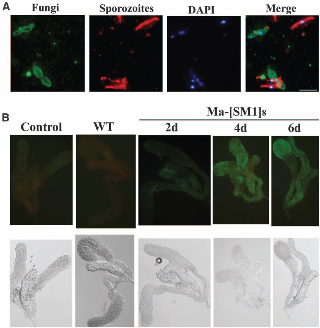
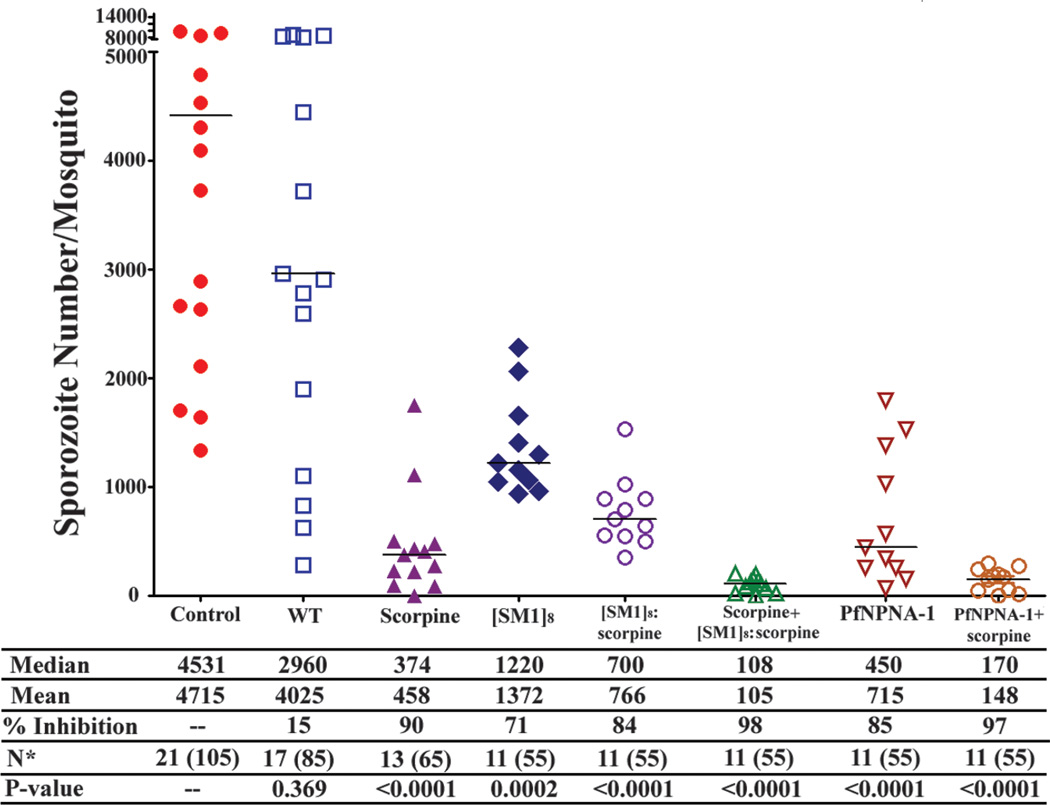
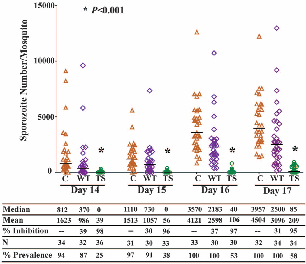
Metarhizium anisopliae infects mosquitoes through the cuticle and proliferates in the hemolymph. To allow M. anisopliae to combat malaria in mosquitoes with advanced malaria infections, we produced recombinant strains expressing molecules that target sporozoites as they travel through the hemolymph to the salivary glands. Eleven days after a Plasmodium-infected blood meal, mosquitoes were treated with M. anisopliae expressing salivary gland and midgut peptide 1 (SM1), which blocks attachment of sporozoites to salivary glands; a single-chain antibody that agglutinates sporozoites; or scorpine, which is an antimicrobial toxin. These reduced sporozoite counts by 71%, 85%, and 90%, respectively. M. anisopliae expressing scorpine and an [SM1](8):scorpine fusion protein reduced sporozoite counts by 98%, suggesting that Metarhizium-mediated inhibition of Plasmodium development could be a powerful weapon for combating malaria.


Figures

Fig. 1
Fig. 1
(A) Coexistence of Metarhizium hyphal bodies and P. falciparum sporozoites in the hemolymph of infected mosquitoes. Hemolymph from mosquitoes 17 days after a Plasmodium-infected blood meal and 6 days after infection with M. anisopliae shows immunostained M. anisopliae (green) and P. falciparum (red). DNA was stained with DAPI (blue, 4´,6´-diamidino-2-phenylindole). Scale bar indicates 10 µm. (B) The timing of expression of [SM1]8 by M. anisopliae–[SM1]8 was estimated by detecting fluorescently immunostained [SM1]8 binding to mosquito salivary glands. (Top row) Salivary glands with fluorescently stained [SM1]8 2, 4, and 6 days postinfection. The intensity of fluorescent signal peaked at 4 days. (Bottom row) Differential interference contrast images of the salivary glands shown in the top row. Control, control salivary glands were from mosquitoes not infected with M. anisopliae; WT, salivary glands from mosquitoes infected with wild-type M. anisopliae.
Fig. 2
Fig. 2
Expression of antiplasmodials by M. anisopliae inhibits sporozoite invasion of salivary glands. Mosquitoes were infected with the wild-type or transgenic Metarhizium strains 11 days after a Plasmodium-infected blood meal, and 6 days later salivary glands from each treatment were dissected, pooled into groups each containing glands from five mosquitoes, and homogenized for sporozoite counting. M. anisopliae strains were engineered to express [SM1]8, PfNPNA-1, scorpine, and an [SM1]8:scorpine fusion protein. Mosquitoes were also co-inoculated with M. anisopliae strains expressing scorpine and an [SM1]8:scorpine fusion protein or with strains expressing scorpine and PfNPNA-1. N, the number of groups (the total number of mosquitoes is shown in parenthesis). Statistical significance was determined by Mann-Whitney test, α = 0.05. The P values given in this figure were calculated by comparing each combination of Plasmodium– and M. anisopliae–infected mosquitoes versus control mosquitoes only infected with Plasmodium. The horizontal lines represent the medians.
Fig. 3
Fig. 3
Sporozoite prevalence and density in mosquitoes infected by transgenic (TS) Metarhizium strains expressing scorpine and [SM1]8:scorpine. Mosquitoes were each infected with ~90 Metarhizium spores 11 days after a Plasmodium-infected blood meal, and salivary glands were scored for sporozoites. Sporozoites were first detected on day 14. The absence of sporozoites determined microscopically was confirmed by PCR (fig. S9). C, control mosquitoes infected with Plasmodium only; WT, mosquitoes infected with Plasmodium and the wild-type M. anisopliae strain. Statistical significance was determined by Mann-Whitney test, α = 0.05. The horizontal lines represent the medians.

Comment in

Similar articles

Cited by

References

    1. World Health Organisation. World Malaria Report. Geneva: World Health Organisation; 2009.
    1. Müller P, Donnelly MJ, Ranson H. BMC Genomics. 2007;8:36. - PMC - PubMed
    1. Knols BG, Bukhari T, Farenhorst M. Future Microbiol. 2010;5:339. - PubMed
    1. Scholte E-J, et al. Science. 2005;308:1641. - PubMed
    1. Thomas MB, Read AF. Nat. Rev. Microbiol. 2007;5:377. - PubMed

Publication types

MeSH terms