. 2011 Feb 25;331(6020):1078-80.
doi: 10.1126/science.1200708.

Transient regenerative potential of the neonatal mouse heart

Affiliations

Transient regenerative potential of the neonatal mouse heart

Enzo R Porrello et al. Science. .

Abstract

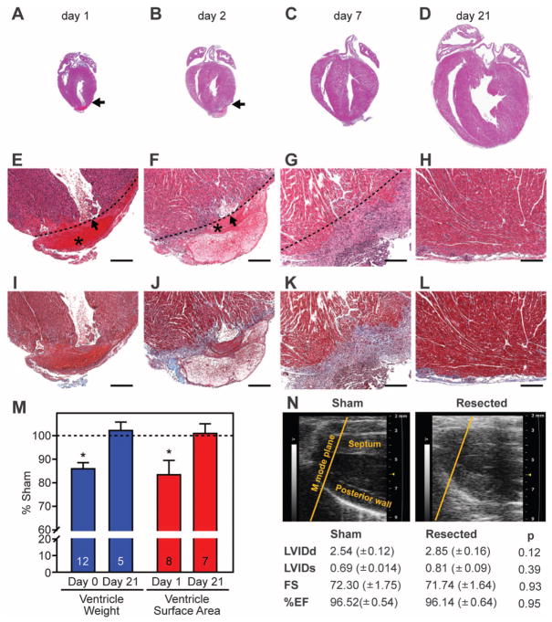
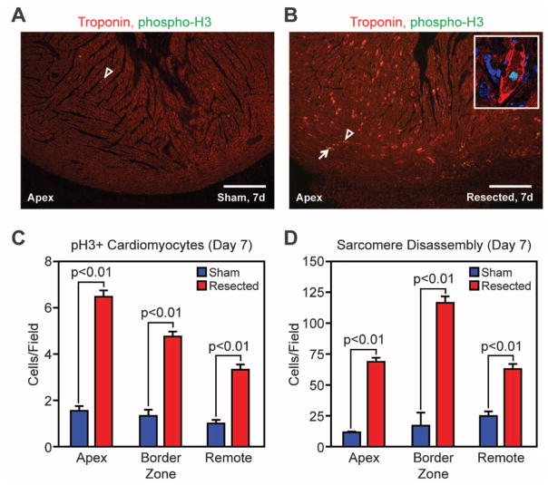
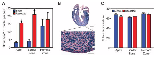
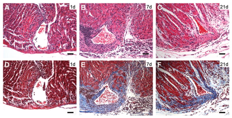
Certain fish and amphibians retain a robust capacity for cardiac regeneration throughout life, but the same is not true of the adult mammalian heart. Whether the capacity for cardiac regeneration is absent in mammals or whether it exists and is switched off early after birth has been unclear. We found that the hearts of 1-day-old neonatal mice can regenerate after partial surgical resection, but this capacity is lost by 7 days of age. This regenerative response in 1-day-old mice was characterized by cardiomyocyte proliferation with minimal hypertrophy or fibrosis, thereby distinguishing it from repair processes. Genetic fate mapping indicated that the majority of cardiomyocytes within the regenerated tissue originated from preexisting cardiomyocytes. Echocardiography performed 2 months after surgery revealed that the regenerated ventricular apex had normal systolic function. Thus, for a brief period after birth, the mammalian heart appears to have the capacity to regenerate.


Figures

Fig. 1
Fig. 1
Regeneration of the ventricular myocardium of neonatal mice (A to D) Hematoxylin and eosin (H&E) staining of the mouse heart at 1, 2, 7, and 21 days post-resection (dpr). Arrow denotes injury site. (E to H) H&E-stained sections at higher magnification. The asterisk marks a large blood clot adjacent to the left ventricular chamber (arrow) at 1 and 2 dpr. Dashed line indicates the resection plane. Scale bars, 200 μm. (I to L) Trichrome-stained serial sections showing early deposition of epicardial extracellular matrix (blue staining) at 7 dpr, with minimal evidence of cardiac fibrosis by day 21. Scale bars, 200 μm. (M) Quantification of surgical reproducibility and regeneration. Ventricle weights and sagittal section surface area are presented as percentages of sham-operated controls. Numbers of samples analyzed are indicated within the bars. Values are presented as means ± SEM; *P < 0.05. (N) Images of 2D echocardiogram in the parasternal long axis view. Yellow line indicates motion (M) mode plane. Lower panel shows left ventricular (LV) dimensions and function of sham-operated and resected hearts at 60 dpr. Left ventricular internal diameter at end systole (LVIDs) and end diastole (LVIDd) were used for calculating fractional shortening (FS) and ejection fraction (EF). Values are presented as means ± SEM; n = 3 per group.
Fig. 2
Fig. 2
Cardiomyocyte proliferation accompanies regeneration of the neonatal mouse heart. (A and B) Cardiomyocyte mitoses were identified by staining for pH3 (green), troponin T (red), and nuclei (blue) in sham-operated (A) and resected (B) hearts. Arrowheads denote cardiomyocytes with disassembled sarcomeres; arrows show cardiomyocytes positive for pH3. Scale bars, 200 μm. The inset is a high-magnification image of a cardiomyocyte with disassembled sarcomeres positively stained for pH3. (C and D) Quantification of pH3 staining (C) and sarcomere disassembly (D) at 7 dpr. Quantitative analysis represents counts per field from four independent samples per group.
Fig. 3
Fig. 3
The majority of newly formed cardiomyocytes within the regenerated apex are derived from preexisting cardiomyocytes. (A) Quantitative analysis of BrdU/Nkx2.5+ nuclei at 21 dpr in sham-operated and resected hearts, after three BrdU pulses (at 1, 7, and 14 dpr). Quantitative analysis represents counting of multiple fields from three independent samples per group (~12 fields per group). Values are presented as means ± SEM; *P < 0.05. (B) β-Galactosidase enzymatic staining of αMHC-MerCreMer; Rosa26-lacZ reporter mouse heart at 21 dpr after a single dose of tamoxifen at birth. Scale bars, 1 mm (upper panel), 100 μm (lower panel). (C) Quantification of the percentage of lacZ-positive myocardium showing no difference between sham-operated and resected hearts at 21 dpr. Quantitative analysis represents counting of multiple fields from three independent samples per group (~18 fields per group). Values are presented as means ± SEM.
Fig. 4
Fig. 4
Lack of regeneration after apical resection of 7-day-old mice. (A to C) H&E staining at 1, 7, and 21 dpr, respectively. (D to F) Trichrome staining at 1, 7, and 21 dpr. Note fibrotic scar (blue staining) surrounding resected ventricular chamber at 7 and 21 dpr [(E) and (F)]. Scale bars, 200 μm.

Similar articles

Cited by

References

    1. Oberpriller JO, Oberpriller JC. J Exp Zool. 1974;187:249. - PubMed
    1. Poss KD, Wilson LG, Keating MT. Science. 2002;298:2188. - PubMed
    1. Jopling C, et al. Nature. 2010;464:606. - PMC - PubMed
    1. Kikuchi K, et al. Nature. 2010;464:601. - PMC - PubMed
    1. Drenckhahn JD, et al. Dev Cell. 2008;15:521. - PubMed

Publication types