Comparative Study
. 2011 Apr 15;108(8):985-95.
doi: 10.1161/CIRCRESAHA.110.233775. Epub 2011 Feb 24.

Human atherosclerotic plaque alternative macrophages display low cholesterol handling but high phagocytosis because of distinct activities of the PPARγ and LXRα pathways

Affiliations
Comparative Study

Human atherosclerotic plaque alternative macrophages display low cholesterol handling but high phagocytosis because of distinct activities of the PPARγ and LXRα pathways

Giulia Chinetti-Gbaguidi et al. Circ Res. .

Abstract

Rationale: A crucial step in atherogenesis is the infiltration of the subendothelial space of large arteries by monocytes where they differentiate into macrophages and transform into lipid-loaded foam cells. Macrophages are heterogeneous cells that adapt their response to environmental cytokines. Th1 cytokines promote monocyte differentiation into M1 macrophages, whereas Th2 cytokines trigger an "alternative" M2 phenotype.

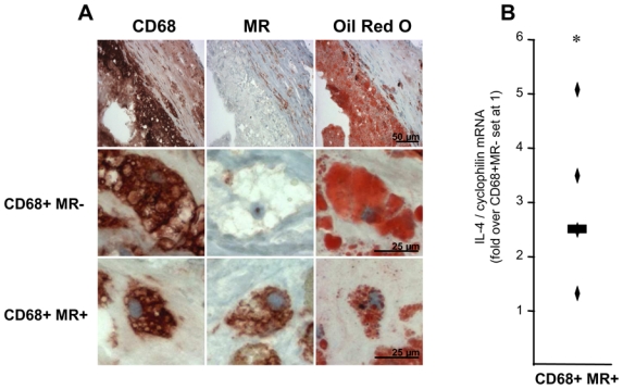
Objective: We previously reported the presence of CD68(+) mannose receptor (MR)(+) M2 macrophages in human atherosclerotic plaques. However, the function of these plaque CD68(+)MR(+) macrophages is still unknown.

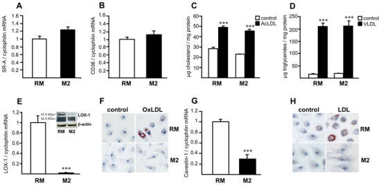
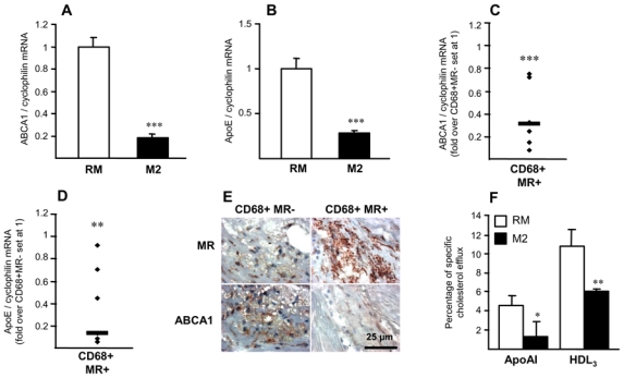
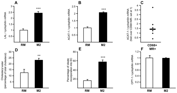
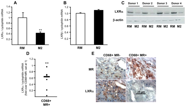
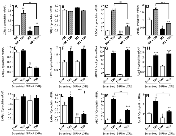
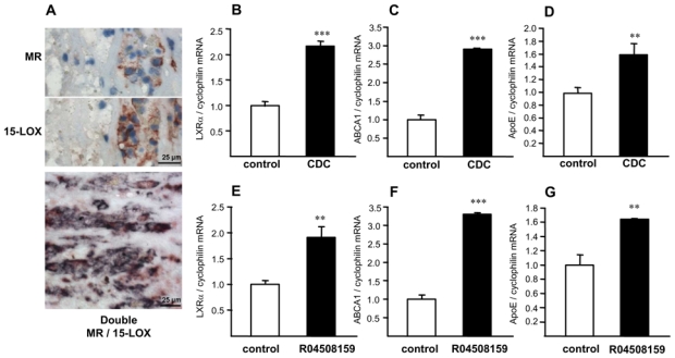
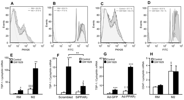
Methods and results: Histological analysis revealed that CD68(+)MR(+) macrophages locate far from the lipid core of the plaque and contain smaller lipid droplets compared to CD68(+)MR(-) macrophages. Interleukin (IL)-4-polarized CD68(+)MR(+) macrophages display a reduced capacity to handle and efflux cellular cholesterol because of low expression levels of the nuclear receptor liver x receptor (LXR)α and its target genes, ABCA1 and apolipoprotein E, attributable to the high 15-lipoxygenase activity in CD68(+)MR(+) macrophages. By contrast, CD68(+)MR(+) macrophages highly express opsonins and receptors involved in phagocytosis, resulting in high phagocytic activity. In M2 macrophages, peroxisome proliferator-activated receptor (PPAR)γ activation enhances the phagocytic but not the cholesterol trafficking pathways.

Conclusions: These data identify a distinct macrophage subpopulation with a low susceptibility to become foam cells but high phagocytic activity resulting from different regulatory activities of the PPARγ-LXRα pathways.


Figures

Figure 1
Figure 1. Identification of distinct macrophage sub-populations in human atherosclerotic plaques
Panel A. Immunostaining (top row) and higher magnification (bottom rows) of representative stainings for CD68, MR and Oil red O in human carotid atherosclerotic lesions. Scale bars are shown. Panel B. Q-PCR analysis of IL-4 performed on RNA from LCM isolated CD68+MR− and CD68+MR+ macrophage-rich areas. mRNA levels were normalized to cyclophilin mRNA and expressed relative to the levels in CD68+MR− area set at 1. Each point corresponds to a single atherosclerotic plaque. The median value is shown. Statistically significant differences are indicated (t-test; *p< 0.05).
Figure 2
Figure 2
Alternative macrophage differentiation decreases native and oxidized LDL accumulation. Panels A,B – E,G. Q-PCR analysis of SR-A (A), CD36 (B), LOX-1 (E) and caveolin-1 (G) mRNA in primary human resting (RM) or alternative macrophages (M2). mRNA levels were normalized to cyclophilin mRNA and results expressed as mean ± SD of triplicate determinations relative to the levels in RM set at 1. Panels C,D. RM or M2 macrophages were loaded with AcLDL (C) or with VLDL (D) and cellular cholesterol or triglycerides determined, respectively. Results are the mean ± SD of triplicate determinations. Statistically significant differences between RM and M2 are indicated (t-test; ***p< 0.001). Panel E. LOX-1 protein expression was determined in RM and M2 macrophages by western blot analysis. Panels F,H. RM or M2 macrophages were incubated with oxidized LDL (OxLDL) (F) or native LDL (LDL) (H) and Oil red O staining performed. Results are representative of 3 independent experiments.
Figure 3
Figure 3. Alternative macrophages display a lower cholesterol efflux capacity
Panels A–D. Q-PCR analysis of ABCA1 (A), ApoE (B) mRNA in RM or M2 macrophages. mRNA levels were normalized to cyclophilin mRNA and results expressed as mean ± SD of triplicate determinations relative to the levels in RM set at 1. Statistically significant differences are indicated (t-test; ***p< 0.001). Q-PCR analysis of ABCA1 (C) and ApoE (D) performed on RNA from LCM-isolated CD68+MR− and CD68+MR+ macrophage-rich areas isolated from 7 samples. mRNA levels were normalized to cyclophilin mRNA and expressed relative to the levels in CD68+MR- area set at 1. Each point corresponds to a single atherosclerotic plaque. The median value is shown. Statistically significant differences are indicated (t-test; **p< 0.01, ***p< 0.001). Panel E. MR and ABCA1 immunostaining performed in human carotid atherosclerotic lesions. Scale bar is shown. Panel F. [3H]-cholesterol-loaded macrophages were incubated with medium with or without apoAI or HDL3 to measure cholesterol efflux Values are expressed as percentage of specific cholesterol efflux and are mean ± SD of 3 independent experiments. Statistically significant differences are indicated (t-test; *p< 0.05, **p< 0.01, ***p< 0.001).
Figure 4
Figure 4. Alternative macrophages display enhanced cholesteryl ester formation capacities
Panels A,B,F. Q-PCR analysis of LAL (A), ACAT-1 (B) and CPT-1 (F) mRNA in RM and M2 macrophages, normalized to cyclophilin mRNA and expressed as mean ± SD relative to RM set at 1 from three independent experiments. Statistically significant differences are indicated (t-test; ***p< 0.001). Panel C. Q-PCR analysis of ACAT-1 performed on RNA from LCM isolated CD68+MR− and CD68+MR+ macrophage-rich areas isolated from 7 samples. mRNA levels were normalized to cyclophilin mRNA and expressed relative to the levels in CD68+MR− area set at 1. Each point corresponds to a single atherosclerotic plaque. Median value is shown. Panel D. RM or M2 macrophages were loaded with [3H]-cholesterol AcLDL and lipids extracted and separated by TLC. Spots corresponding to CE and FC were scraped and radioactivity measured. Results are expressed relative to untreated cells set as 1 as mean ± SD of triplicate determinations obtained from 4 independent macrophage preparations. Panel E. RM or M2 macrophages were cholesterol-loaded with AcLDL and cholesteryl ester formation measured by incubation with [14C]oleic acid. Intracellular lipids were extracted and separated by TLC. Spots corresponding to cholesteryl oleate and oleic acid were scraped and radioactivity measured. Cholesteryl ester formation was calculated as percentage of [C14]oleate incorporated into cholesteryl esters. Results are expressed relative to untreated cells set as 1 as mean ± SD of triplicate determinations obtained from 4 independent macrophage preparations.
Figure 5
Figure 5. Decreased LXRα expression in alternatively differentiated macrophages
Panels A,B. Q-PCR analysis of LXRα (A) and LXRβ (B) in RM and M2 macrophages. mRNA levels were normalized to cyclophilin mRNA and expressed as means ± SD relative to RM set at 1 from three independent experiments. Statistically significant differences are indicated (t-test; *p< 0.05, **p< 0.01). Panel C. LXRα protein expression analyzed by western blot in RM and M2 macrophages isolated from 4 different donors. Panel D. Q-PCR analysis of LXRα performed on RNA from LCM-isolated CD68+MR− and CD68+MR+ macrophage-rich areas isolated from 7 samples. mRNA levels were normalized to cyclophilin mRNA and expressed relative to the levels in CD68+MR− area set at 1. Each point corresponds to a single atherosclerotic plaque. The median value is shown. Statistically significant differences are indicated (t-test; **p< 0.01). Panel E. MR and LXRα immunostaining performed in human carotid atherosclerotic lesions. Scale bar is shown.
Figure 6
Figure 6. Decreased LXRα activity in M2 macrophages
Panels A–D. Q-PCR analysis of LXRα (A), LXRβ (B), ABCA1 (C) and ApoE (D) in RM or M2 macrophages treated or not with T0901317 (T09). mRNA levels were normalized to cyclophilin mRNA and expressed as means ± SD relative to RM set at 1 from three independent experiments. Statistically significant differences are indicated (t-test; RM vs M2 § p< 0.05, §§ p< 0.01, §§§ p< 0.001; and T09 treated vs control **p< 0.01, ***p< 0.001). Panels E–N. Q-PCR analysis of LXRβ, LXRα, ABCA1 and ApoE in M2 macrophages transfected with non-silencing control (scrambled), LXRβ siRNA (E–H) or LXRα siRNA (I–N) and treated or not with T0901317 (T09). Results were normalized to cyclophilin mRNA and expressed relative to the levels in control-siRNA transfected cells set at 1 (mean ± SD of two independent experiments). Statistically significant differences are indicated (t-test; siRNA vs scrambled §p<0.05, §§p<0.01, T09-treated vs control *p<0.05, **p<0.01, ***p<0.001).
Figure 7
Figure 7. 15-LOX inhibition restores the expression of LXRα and its target genes in M2 macrophages
Panel A. MR and 15-LOX immunostaining in human carotid atherosclerotic lesions. Bottom: Double staining of MR (red) and 15-LOX (blue). Most cells present double staining (purple). Scale bar is shown. Panels B–G. Q-PCR analysis of LXRα (B & E), ABCA1 (C & F) and ApoE (D & G) performed in M2 macrophages in the absence or in the presence of the CDC or the R04508159 compounds. mRNA levels were normalized to cyclophilin mRNA and expressed as means ± SD relative to untreated cells set at 1 from three independent experiments. Statistically significant differences are indicated (t-test; **p< 0.01, ***p< 0.001).
Figure 8
Figure 8. Alternative macrophage phagocytic activity is enhanced by PPARγ activation
Panels A,B. Phagocytosis of apoptotic cells (A) and fluorescent beads (B) in RM and M2 macrophages. Panels C,D. Phagocytosis of apoptotic cells (C) and fluorescent beads (D) in M2 macrophages in the absence or in the presence of GW1929 (600 nmol/L) for 24h. White histogram: isotype control. Panels E–H. Q-PCR analysis of TSP-1 (E–G) and CD47 mRNA (H) in RM and M2 macrophages 24h-tretaed with GW1929 (600 nmol/L) (E,H), or in RM macrophages transfected with control or human PPARγ siRNA (F) or infected with Ad-GFP or Ad-PPARγ (G) and subsequently treated or not with GW1929 (600 nmol/L) during 24h. TSP-1 and CD47 mRNA levels were normalized to cyclophilin mRNA and expressed as means ± SD relative to control set at 1, from three independent experiments. Statistically significant differences are indicated (t-test; M2 vs RM, §p<0.05, §§§p<0.001; Ad-PPARγ vs Ad-GFP, ††p<0.01; GW1929-treated vs control, *p<0.05, **p<0.01, ***p<0.001).

Comment in

  • Macrophage subsets in human atherosclerosis.
    Finn AV, Saeed O, Virmani R. Finn AV, et al. Circ Res. 2012 Apr 27;110(9):e64; author reply e65-6. doi: 10.1161/CIRCRESAHA.112.268714. Circ Res. 2012. PMID: 22539759 No abstract available.

References

    1. Wilson HM, Barker RN, Erwig LP. Macrophages: promising targets for the treatment of atherosclerosis. Curr Vasc Pharmacol. 2009;7:234–43. - PubMed
    1. Libby P. Inflammation in atherosclerosis. Nature. 2002;420:868–874. - PubMed
    1. Libby P, Aikawa M, Schonbeck U. Cholesterol and atherosclerosis. Biochimica Biophysica Acta. 2000;1529:299–309. - PubMed
    1. Buhman KF, Accad M, Farese RV. Mammalian acyl-CoA:cholesterol acyltransferases. Biochimica Biophysica Acta. 2000;1529:142–154. - PubMed
    1. Remaley AT, Rust S, Rosier M, Knapper C, Naudin L, Broccardo C, Peterson KM, Koch C, Arnould I, Prades C, Duverger N, Funke H, Assman G, Dinger M, Dean M, Chimini G, Santamarina-Fojo S, Fredrickson DS, Denefle P, Brewer HB., Jr Human ATP-binding cassette transporter 1 (ABC1): genomic organization and identification of the genetic defect in the original tangier disease kindred. Proceedings of the National Academy of Sciences USA. 1999;96:12685–12690. - PMC - PubMed

Publication types

MeSH terms