Visual fields in neuro-ophthalmology
- PMID: 21350279
- PMCID: PMC3116538
- DOI: 10.4103/0301-4738.77013
Visual fields in neuro-ophthalmology
Abstract
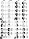
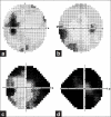
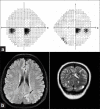
Visual field assessment is important in the evaluation of lesions involving the visual pathways and should be performed at baseline and periodically in the follow-up. Standard automated perimetry has been shown to be adequate in neuro-ophthalmic practise and is now the technique of choice for a majority of practitioners. Goldman kinetic visual fields are useful for patients with severe visual and neurologic deficits and patients with peripheral visual field defects. Visual fields are useful in monitoring progression or recurrence of disease and guide treatment for conditions such as idiopathic intracranial hypertension (IIH), optic neuropathy from multiple sclerosis, pituitary adenomas, and other sellar lesions. They are used as screening tools for toxic optic neuropathy from medications such as ethambutol and vigabatrin. Visual field defects can adversely affect activities of daily living such as personal hygiene, reading, and driving and should be taken into consideration when planning rehabilitation strategies. Visual field testing must be performed in all patients with lesions of the visual pathway.
Conflict of interest statement
Figures


References
-
- Trobe JD, Acosta PC, Krischer JP, Trick GL. Confrontation visual field techniques in the detection of anterior visual pathway lesions. Ann Neurol. 1981;10:28–34. - PubMed
-
- Pandit RJ, Gales K, Griffiths PG. Effectiveness of testing visual fields by confrontation. Lancet. 2001;358:1339–40. - PubMed
-
- Shahinfar S, Johnson LN, Madsen RW. Confrontation visual field loss as a function of decibel sensitivity loss on automated static perimetry. Implications on the accuracy of confrontation visual field testing. Ophthalmology. 1995;102:872–7. - PubMed
Publication types
MeSH terms
Substances
LinkOut - more resources
Full Text Sources

