. 2011 Mar-Apr;59(2):123-36.
doi: 10.4103/0301-4738.77024.

Management of ischemic optic neuropathies

Affiliations

Management of ischemic optic neuropathies

Sohan Singh Hayreh. Indian J Ophthalmol. 2011 Mar-Apr.

Abstract

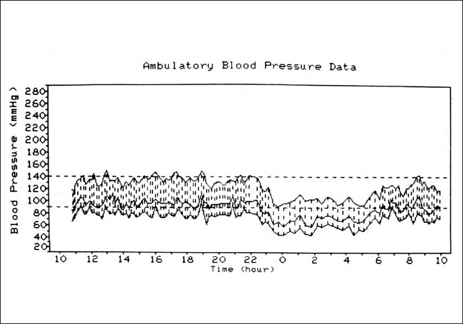
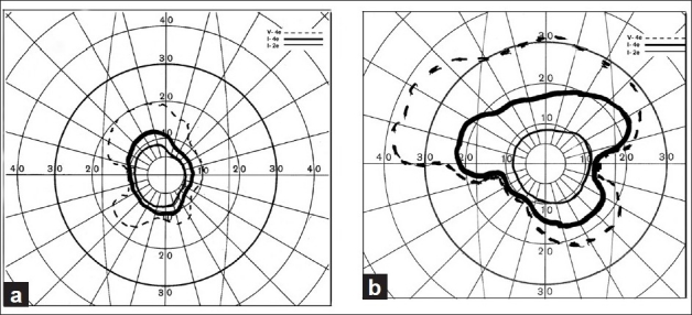
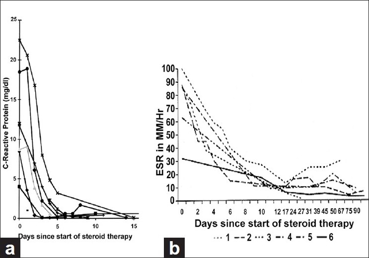
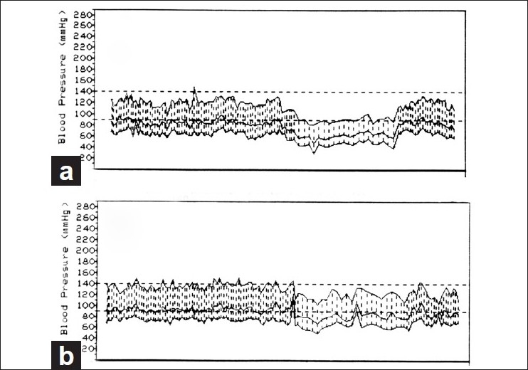
Ischemic optic neuropathies (IONs) consist primarily of two types: anterior ischemic optic neuropathy (AION) and posterior ischemic optic neuropathy (PION). AION comprises arteritic AION (A-AION: due to giant cell arteritis) and non-arteritic AION (NA-AION: due to other causes). PION consists of arteritic PION (A-PION: due to giant cell arteritis), non-arteritic PION (NA-PION: due to other causes), and surgical PION (a complication of several systemic surgical procedures). These five types of ION are distinct clinical entities etiologically, pathogenetically, clinically and from the management point of view. In the management of AION, the first crucial step with patients aged 50 and over is to identify immediately whether it is arteritic or not because A-AION is an ophthalmic emergency and requires urgent treatment with high-dose steroid therapy to prevent any further visual loss in one or both eyes. Patients with NA-AION, when treated with systemic corticosteroid therapy within first 2 weeks of onset, had significantly better visual outcome than untreated ones. Systemic risk factors, particularly nocturnal arterial hypotension, play major roles in the development of NA-AION; management of them is essential in its prevention and management. NA-PION patients, when treated with high-dose systemic steroid therapy during the very early stages of the disease, showed significant improvement in visual acuity and visual fields, compared to untreated eyes. A-PION, like A-AION, requires urgent treatment with high-dose steroid therapy to prevent any further visual loss in one or both eyes. There is no satisfactory treatment for surgical PION, except to take prophylactic measures to prevent its development.


Conflict of interest statement

Conflict of Interest: None declared.

Figures

Figure 1
Figure 1
Schematic representation of blood supply of the optic nerve (A = arachnoid; C = choroid; CRA = central retinal artery; Col. Br. = collateral branches; CRV = central retinal vein; D = dura; LC = lamina cribrosa; ON = optic nerve; P = pia; PCA = posterior ciliary artery; PR = prelaminar region; R = retina, S = sclera; SAS = subarachnoid space)
Figure 2
Figure 2
Ambulatory blood pressure monitoring records (based on individual readings) over a 24-hour period, starting from about 11 a.m., in a 58-year-old woman with bilateral NA-AION and on no medication. The blood pressure is perfectly normal during the waking hours but there is marked nocturnal arterial hypotension during sleep[36]
Figure 3
Figure 3
Fluorescein fundus angiogram of right eye with NA-AION (negative temporal artery biopsy for GCA), showing normal filling of the area supplied by the lateral PCA (including the temporal half of optic disc) but no filling of the area supplied by the medial PCA (including the nasal half of optic disc)[36]
Figure 4
Figure 4
Visual field defects in NA-AION, plotted with Goldmann perimeter (using I-2e, I-4e and V-4e targets). (a) Inferior altitudinal defect with I-2e and inferior nasal defect with I-4e and V-4e; (b) absolute inferior altitudinal defect with I-2e, I-4e and V-4e. The visual acuity in both eyes was 20/20[39]
Figure 5
Figure 5
Fundus photographs of left eye of a 53-year-old man: (a) Normal disc before developing NA-AION, (b) with optic disc edema and hyperemia during the active phase of NA-AION, and (c) after resolution of optic disc edema and development of optic disc pallor (more marked in temporal part than nasal part)[16]
Figure 6
Figure 6
Fundus photograph (a) and fluorescein fundus angiogram (b) of right eye with NA-AION. (a) Optic disc edema, hyperemia and hemorrhages on optic disc; (b) fluorescein fundus angiogram shows non-filling of temporal part of the peripapillary choroid (arrow) and adjacent optic disc and the choroidal watershed zone (arrow)[36]
Figure 7
Figure 7
Right fundus photograph showing optic disc edema and hyperemia, with a splinter hemorrhage (arrow) during the acute phase of NA-AION[16]
Figure 8
Figure 8
Fundus photographs of the both eyes of a 51-year-old diabetic woman, who developed NA-AION, first in the right eye (a, b) and 8 months later in the left eye (c, d). (a, c) Optic disc edema with marked telangiectatic vessels on the optic disc, multiple punctate peripapillary hemorrhages; (b, d) no edema, no abnormal vessels on the disc, and no peripapillary retinal hemorrhages on resolution[41]
Figure 9
Figure 9
Fluorescein fundus angiogram of right eye with NA-AION, showing location of the watershed zone (arrows, WSZ, vertical dark band) in relation to the optic disc. The watershed zone is passing through the temporal part of the disc and adjacent temporal peripapillary choroid[36]
Figure 10
Figure 10
Fundus photograph of right eye with A-AION, showing chalky white optic disc edema during the initial stages[16]
Figure 11
Figure 11
Fundus photographs of right eye with A-AION: (a) Before developing A-AION, (b) 1 week after developing A-AION with chalky white optic disc edema and (c) 4 months later showing optic disc cupping with a cup/disc ratio of 0.8 (note no cup in Fig. 11a before developing A-AION)[16]
Figure 12
Figure 12
Fundus photograph (a) and fluorescein angiogram (b) of left eye with A-AION associated with cilioretinal artery occlusion. (a) Shows chalky white disc edema temporally and retinal infarct in the region of the occluded cilioretinal artery (arrow). (b) Shows normal filling of the area supplied by the lateral PCA, but no filling of the choroid and entire optic disc supplied by the medial PCA or of the cilioretinal artery (arrow)[–18]
Figure 13
Figure 13
Four visual fields in eyes with NA-PION, showing varying sizes and densities of central scotoma and other field defects, with normal peripheral visual fields[13]
Figure 14
Figure 14
Visual fields of (a) right and (b) left eyes with A-PION, showing markedly constricted central visual fields, with complete loss of peripheral fields in both eyes[13]
Figure 15
Figure 15
Graphs of (a) CRP levels and (b) ESR of six patients with GCA, showing their initial responses to high-dose steroid therapy[52]
Figure 16
Figure 16
Two 24-hour ambulatory blood pressure monitoring records (based on individual readings), starting at 10 a.m., of a 63-year old woman taking Verapamil hydrochloride for migraine. Both the records show normal blood pressure during the waking hours. (a) Shows that when she was taking Verapamil at bedtime, during sleep there was a marked degree of nocturnal arterial hypotension (blood pressure falling as low as 80/30 mm Hg). (b) Shows markedly less nocturnal hypotension on stopping the bedtime dose of Verapamil (lowest blood pressure 110/50 mm Hg)

References

    1. Hayreh SS. Anterior ischemic optic neuropathy. Heidelberg: Springer-Verlag; 1975.
    1. Hayreh SS. Blood supply and vascular disorders of the optic nerve. An Inst Barraquer. 1963;4:7–109.
    1. Hayreh SS. The central artery of the retina: Its role in the blood supply of the optic nerve. Br J Ophthalmol. 1963;47:651–63. - PMC - PubMed
    1. Hayreh SS. Blood supply of the optic nerve head and its role in optic atrophy, glaucoma, and oedema of the optic disc. Br J Ophthalmol. 1969;53:721–48. - PMC - PubMed
    1. Hayreh SS. The 1994 Von Sallman lecture. The optic nerve head circulation in health and disease. Exp Eye Res. 1995;61:259–72. - PubMed

MeSH terms