. 2011 Feb 24;470(7335):558-62.
doi: 10.1038/nature09743.

Crystal structure of the CusBA heavy-metal efflux complex of Escherichia coli

Affiliations

Crystal structure of the CusBA heavy-metal efflux complex of Escherichia coli

Chih-Chia Su et al. Nature. .

Abstract

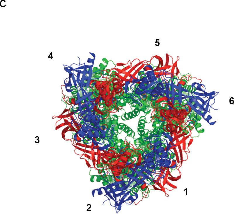
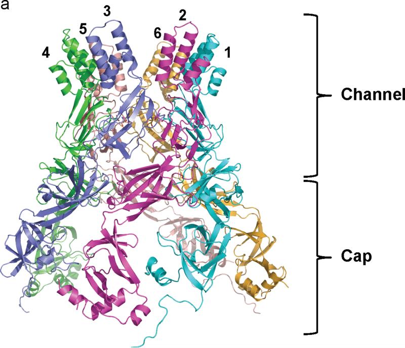
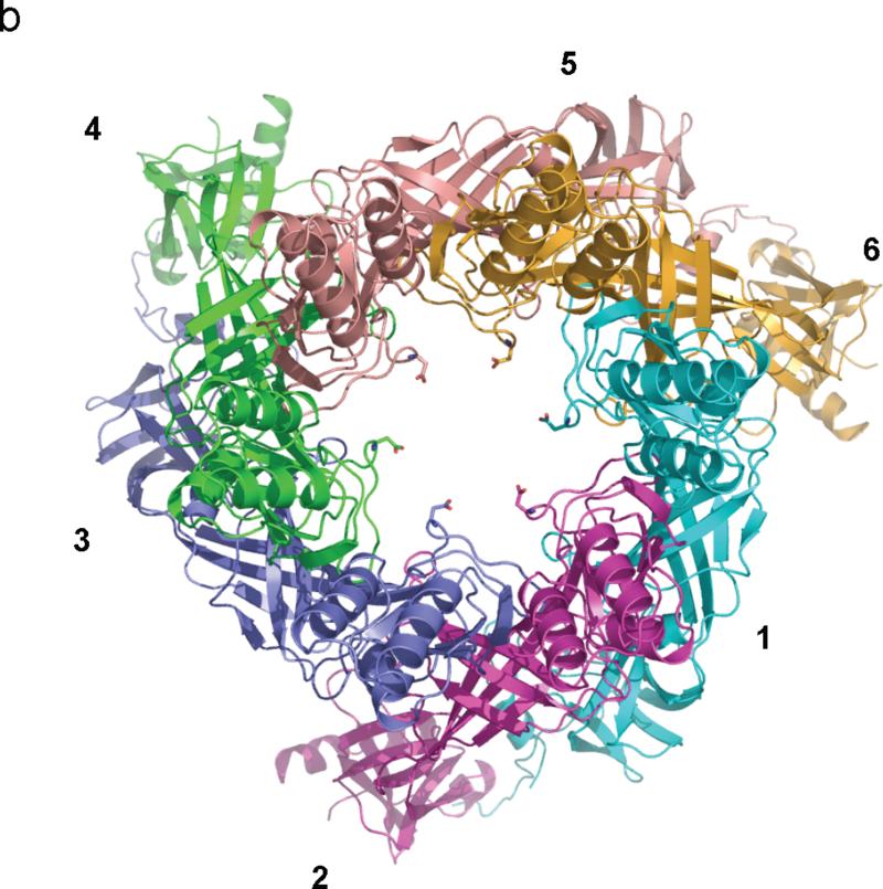
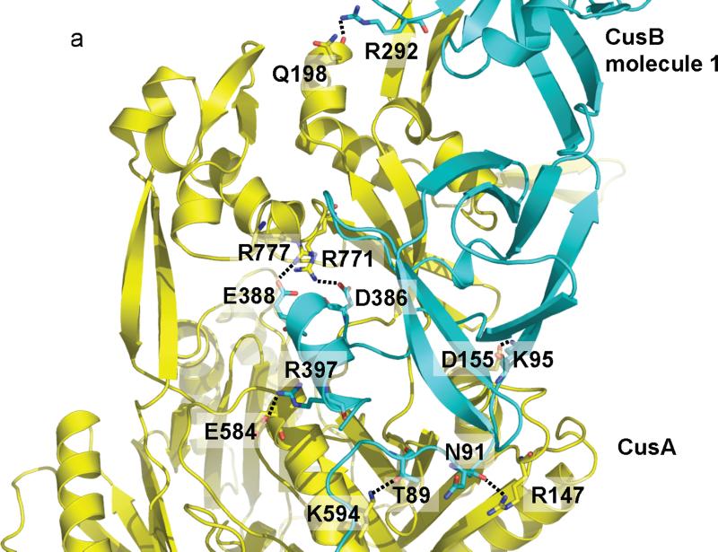
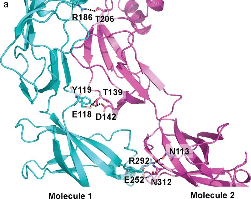
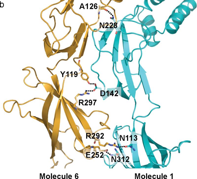
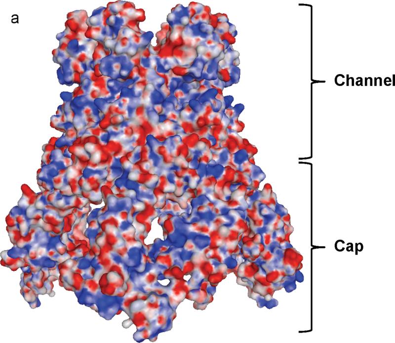
Gram-negative bacteria, such as Escherichia coli, expel toxic chemicals through tripartite efflux pumps that span both the inner and outer membrane. The three parts are an inner membrane, substrate-binding transporter; a membrane fusion protein; and an outer-membrane-anchored channel. The fusion protein connects the transporter to the channel within the periplasmic space. A crystallographic model of this tripartite efflux complex has been unavailable because co-crystallization of the various components of the system has proven to be extremely difficult. We previously described the crystal structures of both the inner membrane transporter CusA and the membrane fusion protein CusB of the CusCBA efflux system of E. coli. Here we report the co-crystal structure of the CusBA efflux complex, showing that the transporter (or pump) CusA, which is present as a trimer, interacts with six CusB protomers and that the periplasmic domain of CusA is involved in these interactions. The six CusB molecules seem to form a continuous channel. The affinity of the CusA and CusB interaction was found to be in the micromolar range. Finally, we have predicted a three-dimensional structure for the trimeric CusC outer membrane channel and developed a model of the tripartite efflux assemblage. This CusC(3)-CusB(6)-CusA(3) model shows a 750-kilodalton efflux complex that spans the entire bacterial cell envelope and exports Cu I and Ag I ions.


Figures

Fig. 1
Fig. 1
Structure of the CusBA efflux complex. (a) Ribbon diagram of the structures of one CusA protomer (green) and two CusB protomers (red and blue) in the asymmetric unit of the crystal lattice. (b) Side view of the CusBA efflux complex. Each subunit of CusA is colored green. Molecules 1, 3 and 5 of CusB are colored red. Molecule 2, 4 and 6 of CusB are in blue. (c) Top view of the CusBA efflux complex. Each subunit of CusA is in green. Molecules 1, 3 and 5 of CusB are in red. Molecules 2, 4 and 6 of CusB are in blue.
Fig. 1
Fig. 1
Structure of the CusBA efflux complex. (a) Ribbon diagram of the structures of one CusA protomer (green) and two CusB protomers (red and blue) in the asymmetric unit of the crystal lattice. (b) Side view of the CusBA efflux complex. Each subunit of CusA is colored green. Molecules 1, 3 and 5 of CusB are colored red. Molecule 2, 4 and 6 of CusB are in blue. (c) Top view of the CusBA efflux complex. Each subunit of CusA is in green. Molecules 1, 3 and 5 of CusB are in red. Molecules 2, 4 and 6 of CusB are in blue.
Fig. 2
Fig. 2
Structure of the hexameric CusB channel. (a) Side view of the hexameric CusB channel. The six molecules CusB are shown in ribbons (cyan, molecule 1; magenta, molecule 2; slate, molecule 3; green, molecule 4; pink, molecule 5; orange, molecule 6). (b) Top view of the hexameric CusB channel. The six molecules CusB are shown in ribbons (cyan, molecule 1; magenta, molecule 2; slate, molecule 3; green, molecule 4; pink, molecule 5; orange, molecule 6). Residue D232, which forms the narrowest region of the central channel, from each subunit of CusB is in sticks. The internal diameter of this narrowest region is ~18 Å.
Fig. 2
Fig. 2
Structure of the hexameric CusB channel. (a) Side view of the hexameric CusB channel. The six molecules CusB are shown in ribbons (cyan, molecule 1; magenta, molecule 2; slate, molecule 3; green, molecule 4; pink, molecule 5; orange, molecule 6). (b) Top view of the hexameric CusB channel. The six molecules CusB are shown in ribbons (cyan, molecule 1; magenta, molecule 2; slate, molecule 3; green, molecule 4; pink, molecule 5; orange, molecule 6). Residue D232, which forms the narrowest region of the central channel, from each subunit of CusB is in sticks. The internal diameter of this narrowest region is ~18 Å.
Fig. 3
Fig. 3
CusA-CusB interactions. (a) The interactions between molecule 1 of CusB and CusA. Residues K95, D386, E388 and R397 of this CusB molecule form four salt bridges with D155, R771, R777 and E584 of CusA, respectively. In addition, T89, the backbone oxygen of N91, and R292 of molecule 1 of CusB form hydrogen bonds with K594, R147, and the backbone oxygen of Q198 of CusA. (b) The interactions between molecule 2 of CusB and CusA. Residues Q108, S109, S253 and N312 of molecule 2 of CusB form hydrogen bonds with Q785, Q194, D800 and Q198 of CusA, respectively. The backbone oxygens of L92 and T335 of this CusB molecule also contribute two additional hydrogen bonds with the side chains of K591 and T808 of the CusA pump to anchor the proteins.
Fig. 3
Fig. 3
CusA-CusB interactions. (a) The interactions between molecule 1 of CusB and CusA. Residues K95, D386, E388 and R397 of this CusB molecule form four salt bridges with D155, R771, R777 and E584 of CusA, respectively. In addition, T89, the backbone oxygen of N91, and R292 of molecule 1 of CusB form hydrogen bonds with K594, R147, and the backbone oxygen of Q198 of CusA. (b) The interactions between molecule 2 of CusB and CusA. Residues Q108, S109, S253 and N312 of molecule 2 of CusB form hydrogen bonds with Q785, Q194, D800 and Q198 of CusA, respectively. The backbone oxygens of L92 and T335 of this CusB molecule also contribute two additional hydrogen bonds with the side chains of K591 and T808 of the CusA pump to anchor the proteins.
Fig. 4
Fig. 4
CusB-CusB interactions. (a) The interactions between molecules 1 and 2 of CusB. Residues E118, Y119, R186, E252 and E292 of molecule 1 of CusB participate to form hydrogen bonds with residues T139, D142, T206, N312 and N113 of molecule 2 of CusB, respectively. These hydrogen-bonded distances are 2.7, 2.7, 3.0, 3.0 and 3.0 Å, respectively. (b) The interactions between molecules 1 and 6 of CusB. Residues N113, N228, and N312 of molecule 1 of CusB pair up with R292, the backbone oxygen of A126, and E252 of molecule 6 of CusB to form three hydrogen bonds. D142 of molecule 1 of CusB also participates to form two hydrogen bonds with Y119 and R297 of molecule 6 of CusB. These hydrogen-bonded distances are 2.7, 2.8, 3.1, 2.7 and 2.8 Å, respectively.
Fig. 4
Fig. 4
CusB-CusB interactions. (a) The interactions between molecules 1 and 2 of CusB. Residues E118, Y119, R186, E252 and E292 of molecule 1 of CusB participate to form hydrogen bonds with residues T139, D142, T206, N312 and N113 of molecule 2 of CusB, respectively. These hydrogen-bonded distances are 2.7, 2.7, 3.0, 3.0 and 3.0 Å, respectively. (b) The interactions between molecules 1 and 6 of CusB. Residues N113, N228, and N312 of molecule 1 of CusB pair up with R292, the backbone oxygen of A126, and E252 of molecule 6 of CusB to form three hydrogen bonds. D142 of molecule 1 of CusB also participates to form two hydrogen bonds with Y119 and R297 of molecule 6 of CusB. These hydrogen-bonded distances are 2.7, 2.8, 3.1, 2.7 and 2.8 Å, respectively.
Fig. 5
Fig. 5
Electrostatic surface potential of CusB. (a) Side view of the electrostatic surface potential of the hexameric CusB channel. This view shows the cap and channel regions formed by the CusB hexamer. Blue (+15 kBT) and red (-15 kBT) indicate the positively and negatively charged areas, respectively, of the protein. (b) Top view of the electrostatic surface potential of the hexameric CusB channel. The widest section of the hexameric channel appears to form at the top edge with its internal diameter of ~56 Å. Blue (+15 kBT) and red (-15 kBT) indicate the positively and negatively charged areas, respectively, of the protein.
Fig. 5
Fig. 5
Electrostatic surface potential of CusB. (a) Side view of the electrostatic surface potential of the hexameric CusB channel. This view shows the cap and channel regions formed by the CusB hexamer. Blue (+15 kBT) and red (-15 kBT) indicate the positively and negatively charged areas, respectively, of the protein. (b) Top view of the electrostatic surface potential of the hexameric CusB channel. The widest section of the hexameric channel appears to form at the top edge with its internal diameter of ~56 Å. Blue (+15 kBT) and red (-15 kBT) indicate the positively and negatively charged areas, respectively, of the protein.

References

    1. Long F, Su C-C, Zimmermann MT, Boyken SE, Rajashankar KR, Jernigan RL, Yu EW. Crystal structures of the CusA heavy-metal efflux pump suggest methionine-mediated metal transport mechanism. Nature. 2010;467:484–488. - PMC - PubMed
    1. Su C-C, Yang F, Long F, Reyon D, Routh MD, Kuo DW, Mokhtari AK, Van Ornam JD, Rabe KL, Hoy JA, Lee YJ, Rajashankar KR, Yu EW. Crystal structure of the membrane fusion protein CusB from Escherichia coli. J. Mol. Biol. 2009;393:342–355. - PMC - PubMed
    1. Franke S, Grass G, Nies DH. The product of the ybdE gene of the Escherichia coli chromosome is involved in detoxification of silver ions. Microbiol. 2001;147:965–972. - PubMed
    1. Franke S, Grass G, Rensing C, Nies DH. Molecular analysis of the copper-transporting efflux system CusCFBA of Escherichia coli. J. Bacteriol. 2003;185:3804–3812. - PMC - PubMed
    1. Tseng TT, Gratwick KS, Kollman J, Park D, Nies DH, Goffeau A, Saier MH., Jr. The RND permease superfamily: an ancient, ubiquitous and diverse family that includes human disease and development protein. J. Mol. Microbiol. Biotechnol. 1999;1:107–125. - PubMed

Publication types

MeSH terms

Associated data