. 2011 Mar;31(3):231-8.
doi: 10.1007/s10059-011-0034-6. Epub 2011 Jan 21.

Targeting the autophagy pathway using ectopic expression of Beclin 1 in combination with rapamycin in drug-resistant v-Ha-ras-transformed NIH 3T3 cells

Affiliations

Targeting the autophagy pathway using ectopic expression of Beclin 1 in combination with rapamycin in drug-resistant v-Ha-ras-transformed NIH 3T3 cells

Ki-Hwan Eum et al. Mol Cells. 2011 Mar.

Abstract

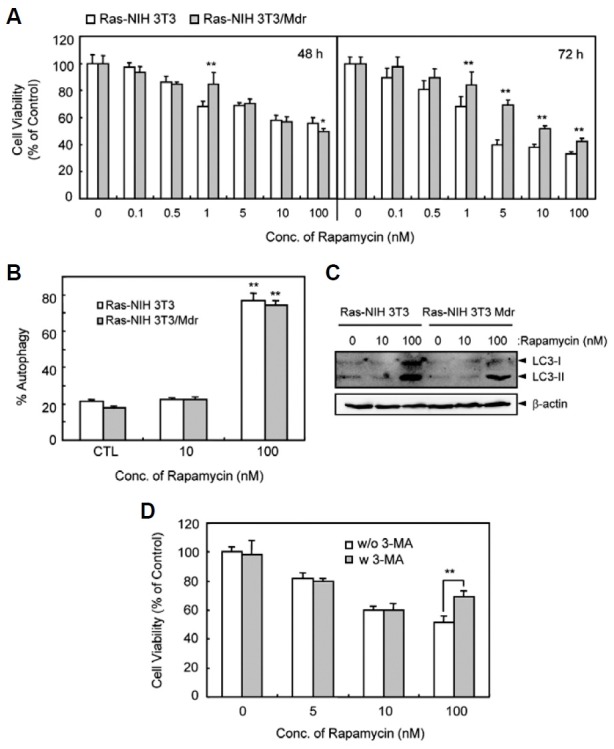
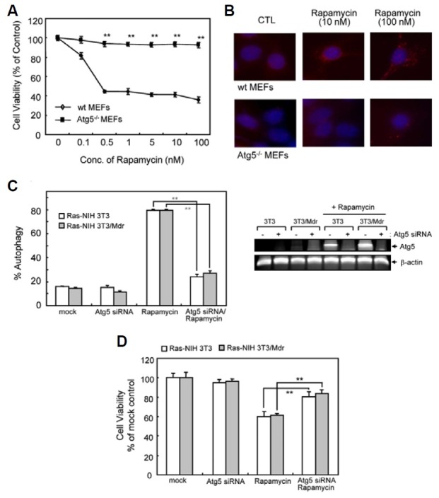
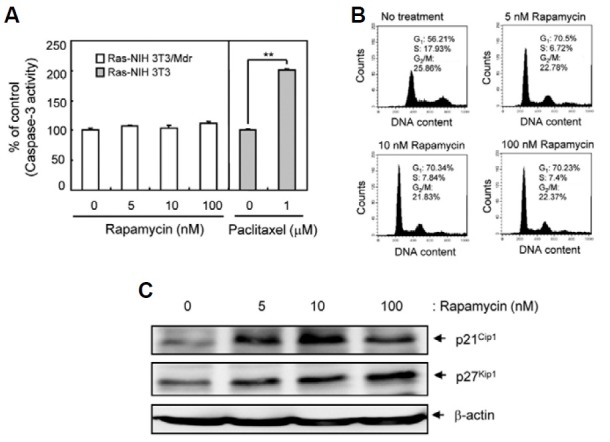
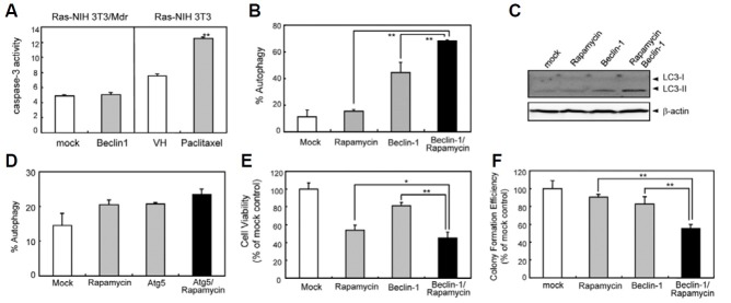
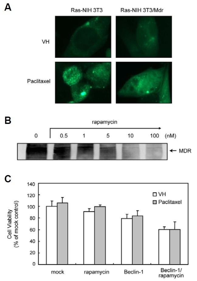
The effectiveness of an apoptosis-targeting therapy may be limited in tumor cells with defects in apoptosis. Recently, considerable attention in the field of cancer therapy has been focused on the mammalian rapamycin target (mTOR), inhibition of which results in autophagic cell death. In our study using multidrug-resistant v-Ha-ras-transformed NIH3T3 (Ras-NIH 3T3/Mdr) cells, we demonstrated that rapamycin-induced cell death may result from 2 different mechanisms. At high rapamycin concentrations (≥ 100 nM), cell death may occur via an autophagy-dependent pathway, whereas at lower concentrations (≤ 10 nM), cell death may occur after G1-phase cell cycle arrest. This effect was accompanied by upregulation of p21(Cip1) and p27(Kip1) expression via an autophagy-independent pathway. We also tested whether inhibition of mTOR with low concentrations of rapamycin and ectopic Beclin-1 expression would further sensitize multidrug resistance (MDR)-positive cancer cells by upregulating autophagy. Rapamycin at low concentrations might be insufficient to initiate autophagosome formation in autophagy but Beclin-1 overexpression triggered additional processes downstream of mTOR during G(1) cell cycle arrest by rapamycin. Our findings suggest that these combination strategies targeting autophagic cell death may yield significant benefits for cancer patients, because lowering rapamycin concentration for cancer treatment minimizes its side effects in patients undergoing chemotherapy.


Figures

Fig. 1.
Fig. 1.. Effect of rapamycin on viability and autophagy of Ras-NIH 3T3 and Ras-NIH 3T3/Mdr cells. (A) Cytotoxic effect of rapamycin. Cells were treated with increasing concentrations of rapamycin ranging from 0.1 to 100 nM for 48 h (left) or for 72 h (right). Cell viability was then evaluated with WST-1 reagent. (B) After 48 h transfection with pEGFP-LC3, cells were incubated for 24 h at 37℃ with rapamycin and immediately analyzed by fluorescence microscopy. To quantify autophagic cells, we counted the number of autophagic cells among 200 GFP-positive cells. *P < 0.01, compared with vehicle control. The results presented are representative of at least three independent exper-iments. (C) The cells were treated with rapamycin as indicated. Electrophoretic mobility change of LC3 from non-autophagic (LC3-I) form to autophagic membrane recruited (LC3-II) form was determined by immunoblotting. (D) Ras- NIH 3T3/Mdr cells were treated with various doses of rapamycin for 24 h in the presence or absence of 1 mM 3-MA and then incubated in 96-well plates for 3 days. Cell growth inhibition was measured by the WST assay. In (A) and (D), the viability of cells treated with vehicle alone was regarded as 100%. Values represent the mean ± SD of quadrupli-cate determinants from one of three representative experiments. **P < 0.01 as determined by Dunnett’s t-test.
Fig. 2.
Fig. 2.. Effect of rapamycin on viability and autophagy of WT and Atg5-/- MEFs. (A) Autophagy-proficient (WT MEFs) and -deficient (Atg5-/- MEFs) cells were treated with increasing concentrations of rapamycin ranging from 0.1 to 100 nM for 72 h. Cell viability was evaluated with WST-1 reagent. The viability of cells treated with vehicle alone was regarded as 100%. Values represent the mean ± SD of quadruplicate determinants from one of three representative experiments. **P < 0.01 as determined by the Dunnett’s T-test as compared to drug-sensitive cells. (B) Autophagy induction was measured by immunofluorescent staining using anti-LC3 (Red). Blue = DAPI staining. (C) Ras-NIH 3T3/Mdr cells were transiently transfected with Atg5 siRNA or scrambled control siRNA for 24 h, together with GFP-LC3. Cells were subsequently washed and then treated with or without rapamycin (10 nM) for 24 h. Autophagy was quantified by the GFP-LC3 puncta. Right inset, RT-PCR showing knock-down of Atg5 mRNA expression by Atg5 siRNA. (D) Atg5 knockdown cells were incubated in the presence or absence of rapamycin in 96-well plates for 3 days. Cell growth inhibition was eva-luated with WST-1 reagent. In (C) and (D), the value of mock-transfected cells was regarded as 100%. Values represent the mean ± SD of quadruplicate determinants from one of three representative experiments. **P < 0.01 as determined by Dunnett’s t-test.
Fig. 3.
Fig. 3.. Detection of G1-phase arrest in Ras-NIH3T3/Mdr cells treated with rapamycin. (A) Pro-apoptotic activity of caspase-3 was determined by detection of the chromophore p-nitroanilide (pNA) after cleavage from the labeled substrate DEVD-pNA in rapamycin- or paclitaxel-treated (positive control) cells. Values represent the mean ± SD of duplicate determinants from one of three representative experiments. **P < 0.01 as determined by the Dunnett’s T-test. (B) Cell cycle progression was assessed by staining fixed cells with propidium iodide to estimate the percentage of cells in the G1 (2N DNA content), G2/M (4N DNA content), and S phases (2 to 4N DNA content). The percentage of cells in each phase of the cell cycle was quantitated using Cell-Quest Pro software and plotted. (C) Whole cell extracts were prepared 48 h post-rapamycin treatment. The expression of p21Cip1 and p27Kip1 were assessed by immuno-blotting. β-actin was assessed as a loading control. The results presented are representative of at least three independent experiments.
Fig. 4.
Fig. 4.. The effect of combined Beclin-1 and rapamycin treatment on autophagy induction and cell growth in Ras-NIH 3T3/Mdr cells. (A) The cells were transiently transfected for 48 h with either a vector expressing full-length Beclin-1 or an empty vector. Ras-NIH 3T3 cells treated with paclitaxel were used as a positive control for apoptosis. Analysis of caspase-3 activity was performed as described in Fig. 2. (B) To quantify the incidence of autophagy, Beclin1-trans-fetced Ras-NIH 3T3/Mdr cells treated with or without rapamycin (10 nM) for 24 h were stained with MDC and visualized by fluorescence microscopy. The autophagic index was calculated as the percentage of MDC-labeled cells out of 200 cells from each treatment group. (C) Beclin1-transfected cells were treated with rapamycin as indicated. Electrophoretic mobility change of LC3 from nonautophagic (LC3-I) form to autophagic membrane recruited (LC3-II) form was determined by immunoblotting. (D) The cells were transiently transfected with either a vector expressing mCherry-Atg5 fusion proteins or an empty vector, together with GFP-LC3 and allowed to express for 24 h. Cells were incubated in the presence or absence of rapamycin (10 nM) for 24 h. To quantify autophagic cells, we counted the number of autophagic cells among 200 GFP-positive cells. (E) Beclin-1-transfetced Ras-NIH 3T3/Mdr cells were incubated in the presence or absence of rapamycin in 96-well plates for 3 days. Cell growth was measured by the WST assay. The viability of cells treated with vehicle alone was regarded as 100%. (F) To identify the effect of rapamycin and Beclin1 when used as single agents or in combination on Ras-NIH 3T3/Mdr cells clonogenic survival, Beclin-1-transfected Ras-NIH 3T3/Mdr cells were incubated in the absence and presence of rapamycin for 7 days. The cells were fixed with methanol and stained with 5% Giemsa solution, and colonies of more than 20 cells were scored. Values represent the mean ± SD of quadruplicate determinants from one of three representative experiments. **P < 0.01; *P < 0.05 as determined by the Dunnett’s t-test.
Fig. 5.
Fig. 5.. The effect of rapamycin on P-glycoprotein in Ras-NIH 3T3/ Mdr cells. (A) Cells were transfected for 48 h with pEGFP-LC3 and then incubated with paclitaxel for 24 h at 37℃. GFP-LC3 distribution was analyzed by fluorescence microscopy. The results presented are representative of at least three independent experiments. (B) The expression of P-glycoprotein was assessed by immunoblotting analysis. Whole cell extracts were prepared 24 h post-rapa-mycin treatment (0.5-100 nM). The expression of P-glycoprotein was assessed by immunoblotting. The results presented are representative of at least three independent experiments (C) Beclin-1-transfected or mock cells were pre-treated with rapamycin for 24 h or vehicle and subsequently treated with or without 1 μM paclitaxel for 2 days. Cell growth was measured by the WST assay. The viability of cells treated with vehicle alone was regarded as 100%. Values represent the mean ± SD of quadruplicate determinants from one of three representative experiments.

References

    1. Arceci R.J., Stieglitz K., Bierer B.E. Immunosuppressants FK506 and rapamycin function as reversal agents of the multidrug resistance phenotype. Blood. (1992);80:1528–1536. - PubMed
    1. Cao Y., Klionsky D.J. Physiological functions of Atg6/ Beclin 1: a unique autophagy-related protein. Cell Res. (2007);17:839–849. - PubMed
    1. Dancey J.E. Inhibitors of the mammalian target of rapamycin. Expert Opin. Investig. Drugs. (2005);14:313–328. - PubMed
    1. Daniel F., Legrand A., Pessayre D. Beclin1 mRNA strongly correlates with Bcl-XL mRNA expression in human hepatocellular carcinoma. Cancer Invest. (2007);25:226–231. - PubMed
    1. Faivre S., Kroemer G., Raymond E. Current development of mTOR inhibitors as anticancer agents. Nat. Rev. Drug Discov. (2006);5:671–688. - PubMed

Publication types

MeSH terms