. 2011 Aug;26(8):1871-82.
doi: 10.1002/jbmr.379.

Periodontal disease and bisphosphonates induce osteonecrosis of the jaws in the rat

Affiliations

Periodontal disease and bisphosphonates induce osteonecrosis of the jaws in the rat

Tara L Aghaloo et al. J Bone Miner Res. 2011 Aug.

Abstract

Bisphosphonates (BPs) are medications used commonly to treat primary and metastatic bone cancer, as well as osteoporosis. Although BPs improve bone mineral density, reduce fracture risk, and reduce hypercalcemia of malignancy, some patients develop BP-related osteonecrosis of the jaws (BRONJ). This devastating complication is defined as clinically exposed bone in the maxillofacial region for more than 8 weeks. Despite an increasing number of BRONJ cases since first reported, the disease pathophysiology remains largely unknown. Since published studies suggest a significant role for dental disease in the pathophysiology of BRONJ, we developed a BRONJ animal model where aggressive periodontal disease is induced by ligature placement around the crown of the right maxillary first molar in the presence of vehicle (veh) or zoledronic acid (ZA), a potent BP. Ligature placement induced significant alveolar bone loss, which was attenuated by ZA treatment. Osteonecrosis was observed associated with ligature-induced periodontitis in the ZA-treated group. This was seen as sequestration and extensive periosteal alveolar bone formation on micro-computed tomography (µCT) in the ligated site of BP-treated animals. Histologic examination confirmed these findings, seen as necrotic bone with diffuse loss of osteocytes and empty lacunae, rimming of the necrotic bone by squamous epithelium and inflammation, and exposure to the oral cavity. Importantly, the rat lesions were strikingly similar to those of BRONJ patients. Our data suggest that dental disease and potent BP therapy are sufficient for BRONJ development in the rat.


Figures

Fig. 1
Fig. 1
(A) Diagram of in vivo periodontal disease model. A sterile ligature was placed subgingivally around the maxillary first molar unilaterally (blue circle) to induce periodontal disease in vehicle- or ZA-treated animals. (B) To quantify ligature-induced alveolar bone loss, the distance between the cementoenamel junction (CEJ) and alveolar crest (AC) in ligated (R) and nonligated (L) sites was measured. Longer distance denotes bone loss. (C) CEJ-AC distance at the distal buccal root of the first molar (D1). (D) CEJ-AC distance at the mesial buccal root of the second molar (M2). *Statistically significantly greater than the nonligated site. #Statistically significantly greater than ZA-treated animals, p<.01.
Fig. 2
Fig. 2
3D μCT reconstructed images of the rat maxilla. (A) Unligated site in vehicle-treated rat. (B) Ligature-induced periodontal disease in vehicle-treated rat. (C, D) Ligature-induced periodontal disease in ZA-treated rat. Blue arrow in panel A points to normal alveolar bone in the interproximal area of the distal root of the first molar and mesial root of the second molar. The yellow arrow in panel B points to periodontal bone loss in vehicle-treated animals at the area of the ligature. In ZA-treated animals, red arrow points to a sequestrum formation (C), and green arrow points to extensive periosteal new bone formation (D) in the area of the ligature.
Fig. 3
Fig. 3
Similarity of rat and human radiographic findings in BRONJ. Sagittal, coronal, and axial CT slices from a rat and a patient with BRONJ demonstrate a bony sequestrum in the alveolar bone (arrows).
Fig. 4
Fig. 4
Similarity of rat and human radiographic findings in BRONJ. Sagittal, coronal, and axial CT slices from a rat and a patient with BRONJ demonstrate significant periosteal bone reaction and expansion of the alveolar ridge dimensions (arrows).
Fig. 5
Fig. 5
Comparison of periosteal bone formation in BRONJ and healthy sites in the rat and patient. To quantify buccal bone thickness in vehicle- versus ZA-treated rats, the buccal width of the alveolar bone was measured at the mesial buccal and distal buccal roots of the first (M1 and D1) and second (M2 and D2) molars in the ligated and nonligated sites on axial μCT slices (A). The measurements on the PD site were calculated as a percentage of the same measurements on the healthy site, and the percent increase was determined (B). To evaluate bone width in BRONJ-involved versus noninvolved sites, cortical bone thickness was measured at the BRONJ site and the same alveolar bone area of the noninvolved site on CBCT images of patients (C). Cortical thickness was expressed as percent thickness of the noninvolved site (D). *Statistically significantly different from the non-PD site, p<.01. #Statistically significantly different from the vehicle- treated group, p<.01. +tatistically significantly different from the noninvolved site, p<.01.
Fig. 6
Fig. 6
Histologic examination of experimentally induced BRONJ. (A, A1) Nonligated site in vehicle-treated animal. (B, B1) Ligature-induced periodontal disease in untreated rat. (C, C1, D, D1) Ligature-induced periodontal disease in ZA-treated rat. (AD: ×10 magnification; A1D1 demonstrate a magnified area of AD.) Black arrows point to viable bone, yellow arrows to necrotic bone, green arrows to inflammatory infiltrate, blue arrows to epithelial rimming of necrotic bone, red arrow to interruption of epithelial continuity, and double white arrow to periosteal bone formation. (Inset: C1, D1) Extensive osteocyte loss with confluent empty lacunae (×40).
Fig. 7
Fig. 7
Rat and human histologic findings in BRONJ. Sections through the jaw of rat (A: ×20; C: ×40) and patient (B: ×20; D: ×40) show osteonecrosis (yellow arrows) with squamous epithelium rimming necrotic bone (blue arrows) and inflammation (green arrows).
Fig. 8
Fig. 8
(A) Number of osteocytic lacunae at the buccal alveolus was measured, and empty lacunae were expressed as a percent of total. (B) Thickness of periosteum at the buccal alveolus was determined. (C) Number of polymorphonuclear neutrophils and (D) intraepithelial lymphocytes was quantified. *Statistically significantly greater than the nonligated site of ZA-treated animals and the nonligated or ligated site of vehicle-treated animals, p<.01. +Statistically significantly greater than the nonligated site, p<.05.
Fig. 9
Fig. 9
×20 TUNEL-stained (A–D) and H&E-stained (A1–D1) adjacent sections from nonligated (A, A1) and ligated (B, B1) sites of a vehicle-treated animal or nonligated (C, C1) and ligated (D, D1) sites of a ZA-treated animal. Several TUNEL+ osteocytes are present in the ZA-treated animal adjacent to an osteonecrotic area (D1). No apoptotic figures are seen in (A–C). (Insets) TUNEL and TUNEL+ cells in detail (×40). (E) Total osteocytes were counted, and percent of TUNEL+ osteocytes was determined. *Statistically significantly greater than the nonligated site of ZA-treated animals and the nonligated or ligated sites of vehicle-treated animals, p < .01.
Fig. 10
Fig. 10
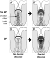
Model of BRONJ pathophysiology. Response of alveolar bone to health (A, C) or dental disease (B, D) in the absence (A, B) or presence (C, D) of BP treatment. BRONJ, depicted by the darker color in bone in panel D requires the presence of both BP and dental disease.

References

    1. Khosla S, Burr D, Cauley J, et al. Bisphosphonate-associated osteonecrosis of the jaw: report of a task force of the American Society for Bone and Mineral Research. J Bone Miner Res. 2007;22:1479–1491. - PubMed
    1. Ruggiero SL, Dodson TB, Assael LA, Landesberg R, Marx RE, Mehrotra B. American Association of Oral and Maxillofacial Surgeons position paper on bisphosphonate-related osteonecrosis of the jaws: 2009 update. J Oral Maxillofac Surg. 2009;67:S2–12. - PubMed
    1. Marx RE, Sawatari Y, Fortin M, Broumand V. Bisphosphonate-induced exposed bone (osteonecrosis/osteopetrosis) of the jaws: risk factors, recognition, prevention, and treatment. J Oral Maxillofac Surg. 2005;63:1567–1575. - PubMed
    1. Marx RE. A new concept in the treatment of osteoradionecrosis. J Oral Maxillofac Surg. 1983;41:351–357. - PubMed
    1. Mehrotra B, Ruggiero S. Bisphosphonate complications including osteonecrosis of the jaw. Hematology, Am Soc Hematol Educ Program. 2006;356–360:515. - PubMed

Publication types

MeSH terms