. 2011 Apr;164(1):90-9.
doi: 10.1111/j.1365-2249.2011.04319.x. Epub 2011 Feb 24.

CD4+ T cells from HIV-1-infected patients recognize wild-type and mutant human immunodeficiency virus-1 protease epitopes

Affiliations

CD4+ T cells from HIV-1-infected patients recognize wild-type and mutant human immunodeficiency virus-1 protease epitopes

N G Muller et al. Clin Exp Immunol. 2011 Apr.

Abstract

Human immunodeficiency virus (HIV)-1 protease is a known target of CD8+ T cell responses, but it is the only HIV-1 protein in which no fully characterized HIV-1 protease CD4 epitopes have been identified to date. We investigated the recognition of HIV-1 protease by CD4+ T cells from 75 HIV-1-infected, protease inhibitor (PI)-treated patients, using the 5,6-carboxyfluorescein diacetate succinimidyl ester-based proliferation assay. In order to identify putative promiscuous CD4+ T cell epitopes, we used the TEPITOPE algorithm to scan the sequence of the HXB2 HIV-1 protease. Protease regions 4-23, 45-64 and 73-95 were identified; 32 sequence variants of the mentioned regions, encoding frequent PI-induced mutations and polymorphisms, were also tested. On average, each peptide bound to five of 15 tested common human leucocyte antigen D-related (HLA-DR) molecules. More than 80% of the patients displayed CD4+ as well as CD8+ T cell recognition of at least one of the protease peptides. All 35 peptides were recognized. The response was not associated with particular HLA-DR or -DQ alleles. Our results thus indicate that protease is a frequent target of CD4+ along with CD8+ proliferative T cell responses by the majority of HIV-1-infected patients under PI therapy. The frequent finding of matching CD4(+) and CD8+ T cell responses to the same peptides may indicate that CD4+ T cells provide cognate T cell help for the maintenance of long-living protease-specific functional CD8+ T cells.


Figures

Fig. 1
Fig. 1
Representative flow analysis using the 5,6-carboxyfluorescein diacetate succinimidyl ester (CFSE) assay (a) and intracellular cytokine staining (ICS) for interleukin (IL)-2 (b). Peripheral blood mononuclear cells (PBMC) from patient 75 were stimulated and stained as described in Materials and methods. (a) Example of a CFSE dilution experiment. PBMC were cultured with peptide PR76–95L89I, a positive control [Streptococcus enterotoxin B (SEB), 1 µg/ml) or no stimulus for 5 days. Cells were acquired on a fluorescence activated cell sorter (FACS)Canto cytometer (Becton Dickinson). At least 5 × 10⁵ events were collected. The boxes show the percentage of CFSElo cells in the CD4+ (left) and CD8+ (right) T cell compartments. (b) ICS experiment with PBMC from the same patient. Cells were cultured with peptide PR76–95L89I, a positive control (SEB, 1 µg/ml) or no stimulus for 12 h with addition of Golgi-stop (Becton Dickinson) after 1 h of incubation. Boxes show the percentage of CD4+ (left) and CD8+ (right) T cells secreting IL-2. Cells were acquired on a FACSCanto cytometer (Becton Dickinson). At least 106 events were collected. All data were analysed using FlowJo™ software (TreeStar).
Fig. 2
Fig. 2
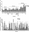
(a) Frequency of human immunodeficiency virus (HIV)-1 infected, protease inhibitor (PI)-treated patients displaying proliferative CD4+ and CD8+ T cell responses against each of the HIV-1 protease peptides. The frequency of responders to each peptide was calculated as follows: (100 × number of patients recognizing the peptide/total number of patients tested). (b) Proportion of HIV-protease peptides recognized by CD4+ and CD8+ T cells from each HIV-1-infected, PI-treated patient. The proportion of peptides recognized by each patient calculated as follows: (100 × number of peptides recognized by the patient/total number of peptides). Peptide-specific CD4+ and CD8+ T cell proliferative responses were assessed by the 5,6-carboxyfluorescein diacetate succinimidyl ester (CFSE)-based antigen-induced proliferation assay as described in Material and methods. Information on the percentage of CFSElo cells in the CD4+ and CD8+ T cell compartments was collected. Peptide-induced T cell responses were displayed as a stimulation index (SI= % CFSElo T cells incubated with peptide/ % CFSElo T cells incubated without peptide). The SI cut-off for CD4+ and CD8+ T cells was 2·5 and 3·2, respectively). Among healthy controls, the median and interquartile ranges of SI in response to all protease peptides were 0·990 (0·923–1·025) for CD4 and 0·945 (0·834–1·040; 25–75%) for CD4+ and CD8+ T cell responses, respectively.
Fig. 3
Fig. 3
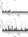
Magnitude of the proliferative CD4+ (a) and CD8+ (b) T cell responses against each human immunodeficiency virus (HIV)-1 protease peptide among protease inhibitor-treated, HIV-1-infected patients undergoing protease inhibitor (PI) therapy. Peptide-specific CD4+ and CD8+ T cell proliferative responses were assessed by the 5,6-carboxyfluorescein diacetate succinimidyl ester (CFSE)-based antigen-induced proliferation assay as described in Material and methods. Information on the percentage of CFSElo cells in the CD4 and CD8 compartments was collected. Peptide-induced T cell responses were quantified as a stimulation index (SI= % CFSElo T cells incubated with peptide/ % CFSElo T cells incubated without peptide). The SI cut-off for CD4+ and CD8+ T cells was 2·5 and 3·2, respectively). Bars represent the median and interquartile ranges of the positive SI. The median and interquartile ranges of background %CFSElo of the tested patients were 0·99 (0·70–1·84) and (0·67 (0·36–1·07) for CD4+ and CD8+ T cells, respectively. Among healthy controls, the median and interquartile ranges of SI in response to all protease peptides were 0·990 (0·923–1·025) for CD4 and 0·945 (0·834–1·040; 25–75%) for CD4+ and CD8+ T cell responses, respectively.

Similar articles

References

    1. Karlsson AC, Deeks SG, Barbour JD, et al. Dual pressure from antiretroviral therapy and cell-mediated immune response on the human immunodeficiency virus type 1 protease gene. J Virol. 2003;77:6743–52. - PMC - PubMed
    1. Mahnke L, Clifford D. Cytotoxic T cell recognition of an HIV-1 reverse transcriptase variant peptide incorporating the K103N drug resistance mutation. AIDS Res Ther. 2006;3:21–4. - PMC - PubMed
    1. Mason RD, Bowmer MI, Howley CM, et al. Antiretroviral drug resistance mutations sustain or enhance CTL recognition of common HIV-1 pol epitopes. J Immunol. 2004;172:7212–19. - PubMed
    1. Mueller SM, Schaetz B, Eismann K, et al. Dual selection pressure by drugs and HLA class I-restricted immune responses on human immunodeficiency virus type 1 protease. J Virol. 2007;81:2887–98. - PMC - PubMed
    1. Samri A, Haas G, Duntze J, et al. Immunogenicity of mutations induced by nucleoside reverse transcriptase inhibitors for human immunodeficiency virus type 1-specific cytotoxic T cells. J Virol. 2000;74:9306–12. - PMC - PubMed

Publication types

MeSH terms