. 2011 May;164(2):256-64.
doi: 10.1111/j.1365-2249.2011.04328.x. Epub 2011 Feb 24.

A soluble thymic stromal lymphopoietin (TSLP) antagonist, TSLPR-immunoglobulin, reduces the severity of allergic disease by regulating pulmonary dendritic cells

Affiliations

A soluble thymic stromal lymphopoietin (TSLP) antagonist, TSLPR-immunoglobulin, reduces the severity of allergic disease by regulating pulmonary dendritic cells

F Zhang et al. Clin Exp Immunol. 2011 May.

Abstract

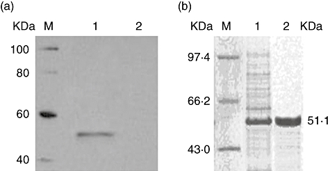
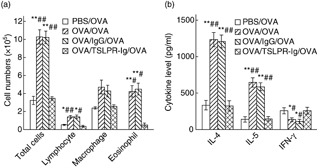
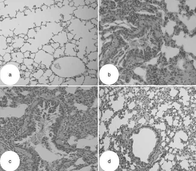
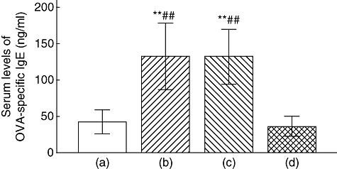
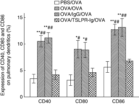
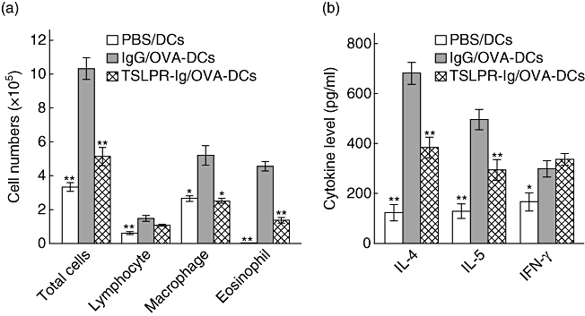
Recent studies show that thymic stromal lymphopoietin (TSLP) plays a critical role in the upstream phase of the allergic cascade to induce T helper type 2 cell (Th2)-dominant allergic diseases. However, the effect of blocking TSLP signalling with the soluble TSLP receptor (TSLPR), TSLPR-immunoglobulin (Ig), on asthma development needs further investigation. Here, we examined the effects of TSLPR-Ig on asthmatic airway inflammation and dendritic cell (DC) function. TSLPR-Ig (comprising the extracellular domain of murine TSLPR and an IgG2a Fc tail) purified from transfected COS-7 cells reduced the expression of CD40, CD80 and CD86 on TSLP-activated DCs in vitro. We also investigated the mechanisms underlying TSLPR-Ig-mediated amelioration of allergic airway inflammation in a murine asthma model. When TSLP signalling was blocked by intratracheal administration of TSLPR-Ig prior to sensitization, allergen-specific serum IgE levels, airway tissue inflammation, inflammatory cell infiltration and Th2 cytokine levels in the bronchiolar lavage fluid (BALF) were reduced significantly. This was because of the TSLP-Ig-mediated down-regulation of co-stimulatory molecule expression on pulmonary DCs. We also transferred bone marrow-derived mature DCs (mDCs) into the airways of asthmatic mice. Intratracheal administration of TSLPR-Ig prior to the transfer of mDCs reduced eosinophilic airway inflammation and Th2 differentiation significantly. Collectively, these data suggest that local use of TSLPR-Ig prevents airway inflammation, at least in part, by regulating DC function, and that blocking TSLP signalling using TSLPR-Ig may be a novel strategy for the treatment of asthma bronchiale.


Figures

Fig. 1
Fig. 1
Expression and purification of the thymic stromal lymphopoietin receptor-immunoglobulin (TSLPR-Ig) fusion protein. (a) Western blot analysis of TSLPR-Ig in the culture medium of transfected COS-7 cells. Lane 1: pTSLPR-Ig transfected COS-7 cells; lane 2: pcDNA-Ig transfected COS-7 cells. M: protein markers. The membrane was then probed with an anti-murine IgG antibody. (b) Sodium dodecyl sulphate-polyacrylamide gel electrophoresis (SDS-PAGE) analysis of purified TSLPR-Ig by Coomassie Blue staining. Soluble TSLPR-Ig was purified from the culture supernatants by protein A Sepharose CL-4B affinity chromatography. Lane 1: total proteins in the culture supernatants from transfected COS-7 cells; lane 2: the purified TSLPR-Ig. M: protein marker.
Fig. 2
Fig. 2
Thymic stromal lymphopoietin receptor-immunoglobulin (TSLPR-Ig) pretreatment suppressed allergic responses. Ovalbumin (OVA)-sensitized and -challenged mice received an intratracheal (i.t.) injection of TSLPR-Ig or control IgG 30 min before each OVA sensitization. (a) Total and differential cells counts and (b) cytokine profiles in the bronchoalveolar lavage fluid (BALF) were analysed 24 h after final OVA exposure. Data represent mean values ± standard error of the mean from six mice per group. *P < 0·05 compared with the phosphate-buffered saline (PBS)/OVA group. **P < 0·01 compared with the PBS/OVA group. #P < 0·05 compared with the OVA/TSLPR-Ig/OVA group. ##P < 0·01 compared with the OVA/TSLPR-Ig/OVA group.
Fig. 3
Fig. 3
Thymic stromal lymphopoietin receptor-immunoglobulin (TSLPR-Ig) pretreatment inhibited lung inflammation. Ovalbumin (OVA)-sensitized and -challenged mice received an intratracheal (i.t.) injection of TSLPR-Ig or control IgG 30 min before each OVA sensitization, and histological examination of the lung tissues (haematoxylin and eosin, ×200 original) was performed 24 h after the final OVA exposure. (a) Phosphate-buffered saline (PBS)/OVA group. (b) OVA/OVA group. (c) OVA/IgG/OVA group. (d) OVA/TSLPR-Ig/OVA group.
Fig. 4
Fig. 4
Thymic stromal lymphopoietin receptor-immunoglobulin (TSLPR-Ig) pretreatment suppressed ovalbumin (OVA)-specific IgE synthesis. Serum levels of OVA-specific IgE in mice from the phosphate-buffered saline (PBS)/OVA group (a), the OVA/OVA group (b), the OVA/IgG/OVA group (c) and the OVA/TSLPR-Ig/OVA group (d), were assessed by enzyme-linked immunosorbent assay. **P < 0·01 compared with group (a). ##P < 0·01 compared with group (d).
Fig. 5
Fig. 5
Thymic stromal lymphopoietin receptor-immunoglobulin (TSLPR-Ig) pretreatment inhibited co-stimulatory molecule expression by pulmonary dendritic cells (DCs). Ovalbumin (OVA)-sensitized and challenged mice received an intratracheal (i.t.) injection of TSLPR-Ig or control IgG 30 min before each OVA sensitization, and the expression of CD40, CD80 and CD86 on pulmonary DCs was analysed by flow cytometry. Data represent mean values ± standard error of the mean from six mice per group. *P < 0·05 compared with the phosphate-buffered saline (PBS)/OVA group. **P < 0·01 compared with the PBS/OVA group. #P < 0·05 compared with the OVA/TSLPR-Ig/OVA group. ##P < 0·01 compared with the OVA/TSLPR-Ig/OVA group.
Fig. 6
Fig. 6
Thymic stromal lymphopoietin receptor-immunoglobulin (TSLPR-Ig) pretreatment impaired the ability of dendritic cells (DCs) to prime T helper type 2 (Th2) responses in vivo. Mice received an intratracheal (i.t.) injection of phosphate-buffered saline (PBS), control IgG or TSLPR-Ig 30 min before i.t. injection of 2 × 10⁶ non-pulsed DCs, or OVA-pulsed DCs (OVA-DCs). They were then exposed to OVA aerosols on days 10–12. (a) Total and differential cells counts and (b) cytokine profiles in the bronchoalveolar lavage fluid (BALF) were assessed 24 h after the final OVA exposure. Data are expressed as mean ± standard error of the mean. *P < 0·05 compared with the IgG/OVA-DCs group. **P < 0·01 compared with the IgG/OVA-DCs group.

References

    1. Komai M, Tanaka H, Masuda T, et al. Role of Th2 responses in the development of allergen-induced airway remodelling in a murine model of allergic asthma. Br J Pharmacol. 2003;138:912–20. - PMC - PubMed
    1. Mazzarella G, Bianco A, Catena E, De Palma R, Abbate GF. Th1/Th2 lymphocyte polarization in asthma. Allergy. 2000;55(Suppl. 61):6–9. - PubMed
    1. Renauld JC. New insights into the role of cytokines in asthma. J Clin Pathol. 2001;54:577–89. - PMC - PubMed
    1. Kuipers H, Lambrecht BN. The interplay of dendritic cells, Th2 cells and regulatory T cells in asthma. Curr Opin Immunol. 2004;16:702–8. - PubMed
    1. Lambrecht BN, Salomon B, Klatzmann D, Pauwels RA. Dendritic cells are required for the development of chronic eosinophilic airway inflammation in response to inhaled antigen in sensitized mice. J Immunol. 1998;160:4090–7. - PubMed

Publication types

MeSH terms