Normalization of proliferation and tight junction formation in bladder epithelial cells from patients with interstitial cystitis/painful bladder syndrome by d-proline and d-pipecolic acid derivatives of antiproliferative factor
- PMID: 21352500
- PMCID: PMC7213761
- DOI: 10.1111/j.1747-0285.2011.01108.x
Normalization of proliferation and tight junction formation in bladder epithelial cells from patients with interstitial cystitis/painful bladder syndrome by d-proline and d-pipecolic acid derivatives of antiproliferative factor
Abstract
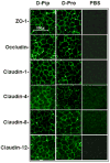
Interstitial cystitis/painful bladder syndrome is a chronic bladder disorder with epithelial thinning or ulceration, pain, urinary frequency and urgency, for which there is no reliably effective therapy. We previously reported that interstitial cystitis/painful bladder syndrome bladder epithelial cells make a glycopeptide antiproliferative factor or 'APF' (Neu5Acα2-3Galβ1-3GalNAcα-O-TVPAAVVVA) that induces abnormalities in normal cells similar to those in interstitial cystitis/painful bladder syndrome cells in vitro, including decreased proliferation, decreased tight junction formation, and increased paracellular permeability. We screened inactive APF derivatives for their ability to block antiproliferative activity of asialylated-APF ('as-APF') in normal bladder cells and determined the ability of as-APF-blocking derivatives to normalize tight junction protein expression, paracellular permeability, and/or proliferation of interstitial cystitis/painful bladder syndrome cells. Only two of these derivatives [Galβ1-3GalNAcα-O-TV-(d-pipecolic acid)-AAVVVA and Galβ1-3GalNAcα-O-TV-(d-proline)-AAVVVA] blocked as-APF antiproliferative activity in normal cells (p < 0.001 for both). Both of these antagonists also 1) significantly increased mRNA expression of ZO-1, occludin, and claudins 1, 4, 8, and 12 in interstitial cystitis/painful bladder syndrome cells by qRT-PCR; 2) normalized interstitial cystitis/painful bladder syndrome epithelial cell tight junction protein expression and tight junction formation by confocal immunofluorescence microscopy; and 3) decreased paracellular permeability of (14) C-mannitol and (3) H-inulin between confluent interstitial cystitis/painful bladder syndrome epithelial cells on Transwell plates, suggesting that these potent APF antagonists may be useful for the development as interstitial cystitis/painful bladder syndrome therapies.
Published 2011. This article is a US Government work and is in the public domain in the USA.
Conflict of interest statement
None of the authors has a conflict of interest related to the work described in this manuscript.
Figures


 ), inactive peptide (
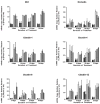
 ), or diluent control (■) twice weekly, and RNA was extracted at 9, 16, 23 and 30 days for qRT-PCR. By day 16, both APF derivatives were able to significantly (p < .05) stimulate tight junction protein expression in IC/PBS cells in vitro resulting in levels similar to those seen in bladder cells from age- and gender-matched normal donors (
 ). PCR was performed in duplicate on three separate occasions for each sample; data are expressed as mean +/- standard error of the mean. (Data shown from experiment with the same IC/PBS cell donor treated simultaneously with either

 ), diluent control (■), or calcium free medium (
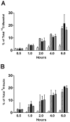
 ) for 16 days prior to incubation with tracer. Data are expressed as mean percent of the radioactivity applied to the apical medium that was recovered in the basal medium at 0.5 hr, 1 hr, 2 hrs, 4 hrs, 6 hrs +/- standard deviation. (Data shown from 4 experiments using cells from 4 different IC/PBS donors).
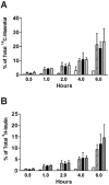
 ), diluent control (■), or calcium free medium (
 ) for 16 days prior to incubation with tracer. Data are expressed as mean percent of the radioactivity applied to the apical medium that was recovered in the basal medium at 0.5 hr, 1 hr, 2 hrs, 4 hrs, 6 hrs +/- standard deviation. (Data shown from 3 experiments using cells from 3 different IC/PBS donors).
Comment in
-
Re: Normalization of proliferation and tight junction formation in bladder epithelial cells from patients with interstitial cystitis/painful bladder syndrome by d-proline and d-pipecolic acid derivatives of antiproliferative factor.J Urol. 2011 Dec;186(6):2264-5. doi: 10.1016/j.juro.2011.07.148. Epub 2011 Oct 26. J Urol. 2011. PMID: 22078590 No abstract available.
Similar articles
-
Regulation of tight junction proteins and bladder epithelial paracellular permeability by an antiproliferative factor from patients with interstitial cystitis.J Urol. 2005 Dec;174(6):2382-7. doi: 10.1097/01.ju.0000180417.11976.99. J Urol. 2005. PMID: 16280852
-
A mouse model for interstitial cystitis/painful bladder syndrome based on APF inhibition of bladder epithelial repair: a pilot study.BMC Urol. 2012 Jun 8;12:17. doi: 10.1186/1471-2490-12-17. BMC Urol. 2012. PMID: 22682521 Free PMC article.
-
The effect of a novel frizzled 8-related antiproliferative factor on in vitro carcinoma and melanoma cell proliferation and invasion.Invest New Drugs. 2012 Oct;30(5):1849-64. doi: 10.1007/s10637-011-9746-x. Epub 2011 Sep 20. Invest New Drugs. 2012. PMID: 21931970 Free PMC article.
-
Cell signaling in interstitial cystitis/painful bladder syndrome.Cell Signal. 2008 Dec;20(12):2174-9. doi: 10.1016/j.cellsig.2008.06.004. Epub 2008 Jun 19. Cell Signal. 2008. PMID: 18602988 Review.
-
Recent developments of intravesical therapy of painful bladder syndrome/interstitial cystitis: a review.Curr Opin Urol. 2006 Jul;16(4):268-72. doi: 10.1097/01.mou.0000232048.81965.16. Curr Opin Urol. 2006. PMID: 16770126 Review.
Cited by
-
Antiproliferative factor signaling and interstitial cystitis/painful bladder syndrome.Int Neurourol J. 2011 Dec;15(4):184-91. doi: 10.5213/inj.2011.15.4.184. Epub 2011 Dec 31. Int Neurourol J. 2011. PMID: 22259731 Free PMC article.
-
Biomarkers in Interstitial Cystitis/Bladder Pain Syndrome with and without Hunner Lesion: A Review and Future Perspectives.Diagnostics (Basel). 2021 Nov 30;11(12):2238. doi: 10.3390/diagnostics11122238. Diagnostics (Basel). 2021. PMID: 34943475 Free PMC article. Review.
-
Glycoamino Acid Analogues of the Thomsen-Friedenreich Tumor-Associated Carbohydrate Antigen: Synthesis and Evaluation of Novel Antiproliferative Factor Glycopeptides.ACS Omega. 2017 Sep 30;2(9):5618-5632. doi: 10.1021/acsomega.7b01018. Epub 2017 Sep 8. ACS Omega. 2017. PMID: 28983523 Free PMC article.
-
Evidence for bladder urothelial pathophysiology in functional bladder disorders.Biomed Res Int. 2014;2014:865463. doi: 10.1155/2014/865463. Epub 2014 May 8. Biomed Res Int. 2014. PMID: 24900993 Free PMC article. Review.
-
Pathomechanism of Interstitial Cystitis/Bladder Pain Syndrome and Mapping the Heterogeneity of Disease.Int Neurourol J. 2016 Nov;20(Suppl 2):S95-104. doi: 10.5213/inj.1632712.356. Epub 2016 Nov 22. Int Neurourol J. 2016. PMID: 27915472 Free PMC article. Review.
References
-
- Johansson SL, Fall M. Clinical features and spectrum of light microscopic changes in interstitial cystitis. J Urol. 1990;143:1118–1124. - PubMed
-
- Skoluda D, Wegner K, Lemmel EM. Critical Notes: Respective immune pathogenesis of interstitial cystitis. (article in German) Urologe A. 1974;13:15–23. - PubMed
-
- Tomaszewski JE, Landis JR, Russack V, Williams TM, Wang LP, Hardy C, Brensinger C, Matthews YL, Abele ST, Kusek JW, Nyberg LM. Biopsy features are associated with primary symptoms in interstitial cystitis: results from the Interstitial Cystitis Database Study Group. Urology. 2001;57:67–81. - PubMed
-
- Lavelle J, Meyers S, Ramage R, Bastacky S, Doty D, Apodaca G, Zeidel ML. Bladder permeability barrier: recovery from selective injury of surface epithelial cells. Am J Physiol Renal Physiol. 2002;283:F242–F253. - PubMed
-
- Acharya P, Beckel J, Ruiz WG, Wang E, Rojas R, Birder L, Apodaca G. Distribution of the tight junction proteins ZO-1, occludin, and claudin-4, -8, and -12 in bladder epithelium. Am J Physiol. 2004;287:F305–F318. - PubMed
Publication types
MeSH terms
Substances
Grants and funding
LinkOut - more resources
Full Text Sources
Medical

