. 2011 Feb 25:11:43.
doi: 10.1186/1471-2180-11-43.

Biochemical and physiological characterization of the GTP-binding protein Obg of Mycobacterium tuberculosis

Affiliations

Biochemical and physiological characterization of the GTP-binding protein Obg of Mycobacterium tuberculosis

Smitha J Sasindran et al. BMC Microbiol. .

Abstract

Background: Obg is a highly conserved GTP-binding protein that has homologues in bacteria, archaea and eukaryotes. In bacteria, Obg proteins are essential for growth, and they participate in spore formation, stress adaptation, ribosome assembly and chromosomal partitioning. This study was undertaken to investigate the biochemical and physiological characteristics of Obg in Mycobacterium tuberculosis, which causes tuberculosis in humans.

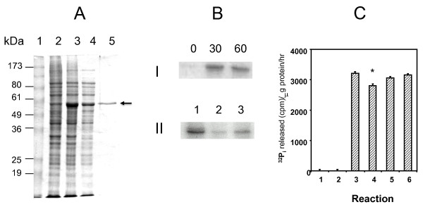
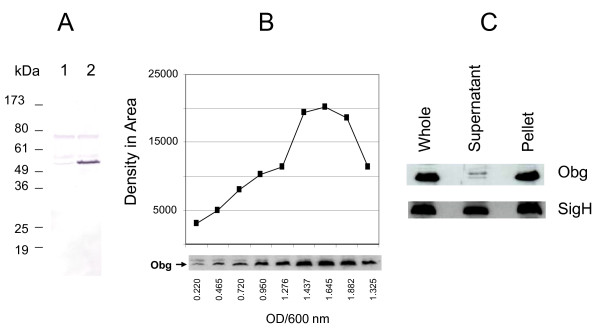
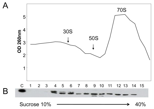
Results: We overexpressed M. tuberculosis Obg in Escherichia coli and then purified the protein. This protein binds to, hydrolyzes and is phosphorylated with GTP. An anti-Obg antiserum, raised against the purified Obg, detects a 55 kDa protein in immunoblots of M. tuberculosis extracts. Immunoblotting also discloses that cultured M. tuberculosis cells contain increased amounts of Obg in the late log phase and in the stationary phase. Obg is also associated with ribosomes in M. tuberculosis, and it is distributed to all three ribosomal fractions (30 S, 50 S and 70 S). Finally, yeast two-hybrid analysis reveals that Obg interacts with the stress protein UsfX, indicating that M. tuberculosis Obg, like other bacterial Obgs, is a stress related protein.

Conclusions: Although its GTP-hydrolyzing and phosphorylating activities resemble those of other bacterial Obg homologues, M. tuberculosis Obg differs from them in these respects: (a) preferential association with the bacterial membrane; (b) association with all three ribosomal subunits, and (c) binding to the stress protein UsfX, rather than to RelA. Generation of mutant alleles of Obg of M. tuberculosis, and their characterization in vivo, may provide additional insights regarding its role in this important human pathogen.


Figures

Figure 1
Figure 1
Analysis of overexpressed Obg and its GTP binding and hydrolysis activities. A. SDS-PAGE protein profile showing overexpression and purification of M. tuberculosis Obg. E. coli was grown in LB broth at 37°C, and lysates were prepared by sonication. Lane 1, Molecular markers; Lanes 2 and 3, extracts of E. coli strain BL21 carrying the overexpression plasmid pTBOBGE in the absence (Lane 2) and presence (Lane 3) of 1 mM IPTG; Lane 4, supernatant of E. coli lysate after 10,000 g centrifugation; Lane 5, His10-Obg after Ni-NTA affinity chromatography. The arrow points to the His10-Obg band. B. Autoradiogram of SDS-PAGE-separated M. tuberculosis His10-Obg after UV-crosslinking with [α32P]GTP. UV-cross-linking was performed by incubating 5 μg of His10-Obg with 10 μCi of [α32P]GTP in the binding buffer as described in the Methods section I. Crosslinking of His10-Obg with [α32P]GTP after 0, 30 and 60 minutes of exposure to UV light (256 nm). II. Crosslinking of His10-Obg with [α32P]GTP for 30 min without any additional GTP or ATP in the reaction mixture (Lane 1) or with 5 mM of unlabeled GTP (Lane 2), or with 500 mM of unlabeled ATP (Lane 3). C. GTPase activity of His10-Obg. GTP hydrolysis of His10-Obg was performed using [γ-32P] GTP at 37°C. The GTPase activity is expressed as 32Pi released (cpm)/μg protein/hour. Columns indicate GTPase activity in the absence of [γ-32P]GTP and His10-Obg (Column 1), in the presence of His10-Obg alone (Column 2), in the presence of both [γ-32P]GTP and His10-Obg (Column 3), in the presence of [γ -32P]GTP, His10-Obg and 5 mM unlabeled GTP (Column 4), in the presence of [γ -32P]GTP, His10-Obg and 5 mM unlabeled GDP (Column 5) and in the presence of [γ-32P]GTP, His10-Obg and 5 mM unlabeled ATP (Column 6). * indicates value significant from column 3 (paired t-test P = 0.0163).
Figure 2
Figure 2
Autoradiogram of SDS-PAGE-separated M. tuberculosis His10-Obg after autophosphorylation. Autophosphorylation reactions were set up by incubating 5 μg of His10-Obg with 10 μCi of [γ-32P] GTP in autophosphorylation buffer, as detailed in the Methods section. A. Autophosphorylation of His10-Obg by [γ-32P] GTP or [γ-32P]ATP after 0, 15, 30 and 60 minutes of incubation at 37°C. B. Autophosphorylation of His10-Obg by [γ-32P]GTP in the presence (+ lane) and absence of (- lane) 1.5 mM MgCl2 . C. Autophosphorylation of His10-Obg by [γ-32P]GTP in the presence of 5 mM (Lane 1), 50 mM (Lane 2) and 500 mM (Lane 3) ATP; 5 mM (Lane 1), 50 mM (Lane 2) and 500 mM (Lane 3) of GTP; 5 mM (Lane 1), 50 mM (Lane 2) and 500 mM (Lane 3) of GDP.
Figure 3
Figure 3
Immunoblot analysis of Obg of M. tuberculosis. A. Immunoblot analysis of Obg from M. tuberculosis strains harboring plasmids. M. tuberculosis strains were grown in 7H9-OADC-TW broth at 37°C to early log phase and lysates prepared using a bead beater and separated (100 μg protein for each lane) on SDS-PAGE. The immunoblots were probed with anti-Obg antiserum (1:500 dilution) followed by alkaline phosphatase labeled anti-rabbit IgG (1:1000 dilution, Zymed). The antibody-incubated blots were then developed with NBT/BCIP substrates. Lane 1, M. tuberculosis carrying the plasmid pMV261(empty vector control); Lane 2, M. tuberculosis carrying the plasmid pMVOBG (plasmid overexpressing Obg). B. Immunoblot analysis of Obg at different growth points in M. tuberculosis culture. Wild type M. tuberculosis was grown in 7H9-OADC-TW broth at 37°C. Lysates were prepared from wild-type M. tuberculosis grown to different ODs at 600 nm, separated (200 μg protein for each lane) on SDS-PAGE, and probed with anti-Obg antiserum (1:500 dilution) followed by peroxidase-labeled anti-rabbit IgG (1:10,000 dilution, Sigma). The blots were developed with an ECL kit (Amersham) and autoradiographed. "Obg" indicates the Obg protein reacting with anti-Obg antiserum. Values below each band indicate the OD value at 600 nm at the time of harvest. The graph above the bands gives the levels of Obg, based on density of the bands using Image J software. C. Immunoblots of Obg in separated soluble vs membrane fractions of M. tuberculosis lysates. The bacteria were grown in 7H9-OADC-TW broth at 37°C to mid-log phase. Lysates were prepared using a bead beater, and the soluble and pellet fractions separated by centrifugation. The protein fractions (200 μg protein for each lane) were separated by SDS-PAGE, blotted and probed with anti-Obg antiserum (1:500 dilution) (marked as Obg) or anti-SigH antiserum (1:1000 dilution) (marked as SigH), followed by peroxidase-labeled anti-rabbit IgG (1:10,000 dilution, Sigma). The blots were developed with an ECL kit (Amersham) and autoradiographed. In the figure, lanes labeled Whole, Supernatant and Pellet represent extracts of whole M. tuberculosis, of the 49,000 g supernatant, and of the 49,000 g pellet, respectively.
Figure 4
Figure 4
Obg cofractionation with ribosomal subunits. M. tuberculosis was grown in 7H9-OADC-TW broth at 37°C, and lysates prepared using a bead beater. About 500 g protein was separated in 10-40% sucrose gradient. A. The ODs of the separated fractions were measured (manually) at 260 nm. B. The proteins in the fractions were then precipitated with ethanol and separated on SDS-PAGE, transferred to nitrocellulose membranes, and probed with anti-Obg antiserum (1:500 dilution), followed by peroxidase-labeled anti-rabbit IgG (1:10,000 dilution, Sigma). The blots were developed with an ECL kit (Amersham) and autoradiographed. Lane C is a whole-cell extract from M. tuberculosis. Lanes 1-15 represent fractions from the top (10% sucrose) to the bottom (40% sucrose) of the sucrose gradient. Fraction 16 was not analyzed in immunoblot.
Figure 5
Figure 5
Growth of M. tuberculosis strains at different time points. M. tuberculosis was grown in 7H9-OADC-TW broth at 37°C. Growth was followed by measuring the OD at 600 nm using 1 ml aliquots. Closed circles: M. tuberculosis carrying the plasmid pMV261 (empty vector control); squares: M. tuberculosis carrying the plasmid pMVOBG (plasmid overexpressing Obg). The data shown are representative findings from three different. experiments.

Similar articles

Cited by

References

    1. Bourne HR, Sanders DA, McCormick F. The GTPase superfamily: a conserved switch for diverse cell functions. Nature. 1990;348(6297):125–132. doi: 10.1038/348125a0. - DOI - PubMed
    1. Kaziro Y, Itoh H, Kozasa T, Nakafuku M, Satoh T. Structure and function of signal-transducing GTP-binding proteins. Annu Rev Biochem. 1991;60:349–400. doi: 10.1146/annurev.bi.60.070191.002025. - DOI - PubMed
    1. Bourne HR, Sanders DA, McCormick F. The GTPase superfamily: conserved structure and molecular mechanism. Nature. 1991;349(6305):117–127. doi: 10.1038/349117a0. - DOI - PubMed
    1. Sprang SR. G protein mechanisms: insights from structural analysis. Annu Rev Biochem. 1997;66:639–678. doi: 10.1146/annurev.biochem.66.1.639. - DOI - PubMed
    1. Pandit SB, Srinivasan N. Survey for g-proteins in the prokaryotic genomes: prediction of functional roles based on classification. Proteins. 2003;52:585–597. doi: 10.1002/prot.10420. - DOI - PubMed

Publication types

MeSH terms

LinkOut - more resources