. 2011 Feb 27:11:5.
doi: 10.1186/1471-5945-11-5.

Targeting of human interleukin-12B by small hairpin RNAs in xenografted psoriatic skin

Affiliations

Targeting of human interleukin-12B by small hairpin RNAs in xenografted psoriatic skin

Rasmus O Bak et al. BMC Dermatol. .

Abstract

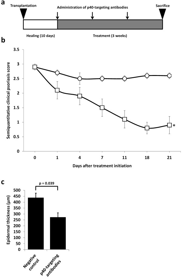
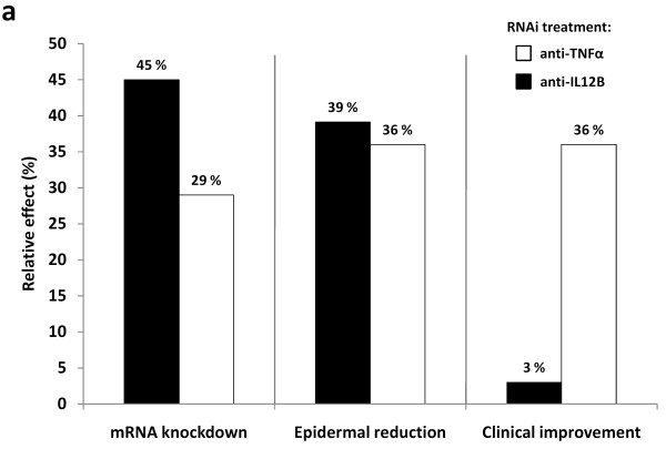
Background: Psoriasis is a chronic inflammatory skin disorder that shows as erythematous and scaly lesions. The pathogenesis of psoriasis is driven by a dysregulation of the immune system which leads to an altered cytokine production. Proinflammatory cytokines that are up-regulated in psoriasis include tumor necrosis factor alpha (TNFα), interleukin-12 (IL-12), and IL-23 for which monoclonal antibodies have already been approved for clinical use. We have previously documented the therapeutic applicability of targeting TNFα mRNA for RNA interference-mediated down-regulation by anti-TNFα small hairpin RNAs (shRNAs) delivered by lentiviral vectors to xenografted psoriatic skin. The present report aims at targeting mRNA encoding the shared p40 subunit (IL-12B) of IL-12 and IL-23 by cellular transduction with lentiviral vectors encoding anti-IL12B shRNAs.

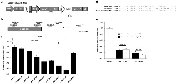
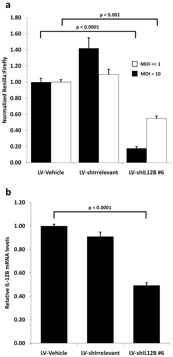
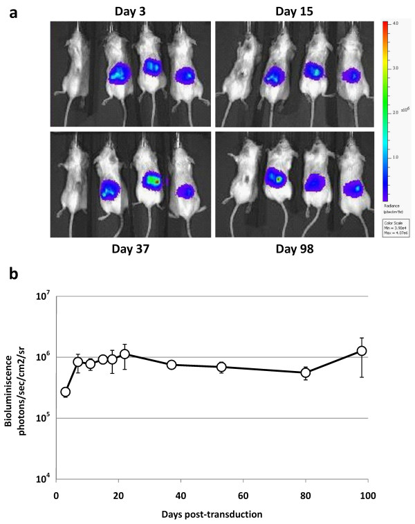
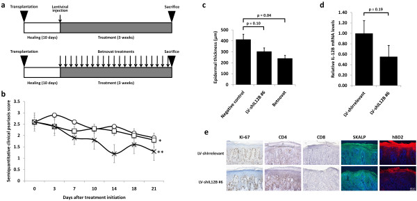
Methods: Effective anti-IL12B shRNAs are identified among a panel of shRNAs by potency measurements in cultured cells. The efficiency and persistency of lentiviral gene delivery to xenografted human skin are investigated by bioluminescence analysis of skin treated with lentiviral vectors encoding the luciferase gene. shRNA-expressing lentiviral vectors are intradermally injected in xenografted psoriatic skin and the effects of the treatment evaluated by clinical psoriasis scoring, by measurements of epidermal thickness, and IL-12B mRNA levels.

Results: Potent and persistent transgene expression following a single intradermal injection of lentiviral vectors in xenografted human skin is reported. Stable IL-12B mRNA knockdown and reduced epidermal thickness are achieved three weeks after treatment of xenografted psoriatic skin with lentivirus-encoded anti-IL12B shRNAs. These findings mimic the results obtained with anti-TNFα shRNAs but, in contrast to anti-TNFα treatment, anti-IL12B shRNAs do not ameliorate the psoriatic phenotype as evaluated by semi-quantitative clinical scoring and by immunohistological examination.

Conclusions: Our studies consolidate the properties of lentiviral vectors as a tool for potent gene delivery and for evaluation of mRNA targets for anti-inflammatory therapy. However, in contrast to local anti-TNFα treatment, the therapeutic potential of targeting IL-12B at the RNA level in psoriasis is questioned.


Figures

Figure 1
Figure 1
Amelioration of psoriasis by treatment of mice xenotransplanted with psoriatic skin with systemically administered p40-targeting antibodies. (a) Schematic schedule of treatment with p40-targeting antibodies (Ustekinumab). Psoriatic skin grafts were xenotransplanted onto the back of SCID mice and allowed to heal for ten days. The mice were then treated by weekly intraperitoneal injection of p40-targeting antibodies or negative control. The mice were sacrificed after three weeks of treatment. (b) Semiquantitative clinical psoriasis scores were given twice weekly for the three-week treatment duration to mice treated with the negative control (open circles, n = 11) or p40-targeting antibodies (open squares, n = 4). Injections were performed at day 0, 7, and 14. All data points are presented as mean ± SEM. *p = 0.003. (c) At treatment endpoint, mice were sacrificed and biopsies from the skin grafts were fixed, paraffin-embedded, H&E-stained, and epidermal thickness was measured in each graft. All data points are presented as mean + SEM.
Figure 2
Figure 2
Development of a lentiviral vector for an easy one-step shRNA oligonucleotide cloning procedure and potency screening of a panel of shRNAs targeting human IL-12B. (a) Schematic overview of the lentiviral vector, pCCL-PGK-Puro-H1-MCS, for an easy one-step shRNA cloning procedure. shRNA oligonucleotides with compatible overhangs can be cloned into the multiple cloning site (MCS) from which shRNA expression will be driven by the H1 promoter. (b) Schematic overview of IL-12B mRNA and the target sites for the seven designed shRNAs. (c) Potency screening of seven shRNAs targeting IL-12B mRNA using the dual luciferase assay. HEK293 cells were co-transfected with the shRNA-encoding lentiviral vector and the psiCHECK-IL12B vector encoding firefly luciferase for transfection normalization and a fusion mRNA consisting of renilla luciferase and IL-12B. Luciferase activities were measured forty-eight hours post-transfection and renilla luciferase activity was normalized to firefly luciferase activity and depicted relative to transfection with the empty lentiviral vector, pCCL-PGK-Puro-H1-MCS, not encoding an shRNA (vehicle). (d) Sequence comparison of the 3' end of the H1 promoter depicting sequence differences introduced in the H1 promoter for cloning of shRNA oligonucleotides. The TATA box is depicted in bold, and sequence differences are underlined. Furthermore, the termination signal of the H1 promoter consists of 5 thymines. (e) Confirmation of full promoter activity from the H1 promoter of pCCL-PGK-Puro-H1-MCS modified to encompass a multiple cloning site for easy one-step cloning of shRNA oligonucleotides. shRNA potency was evaluated when expressed from the two sequence contexts of the H1 promoter, namely the context described by Raoul et. al. (black bars) and the new sequence context of the pCCL-PGK-Puro-H1-MCS (white bars). Comparison of shRNA potency was performed using the dual luciferase assay as described above. All dual luciferase assay experiments were performed at least in triplicates and data are depicted as mean + SEM.
Figure 3
Figure 3
Confirmation of shRNA potency after lentiviral delivery. (a) shRNA potency evaluation after transduction with shRNA-encoding lentiviral vectors. HEK293 cells were transduced at an MOI of 10 (black bars) or << 1 (white bars) followed by puromycin selection for ten days to ensure that cells harbored only a single lentiviral insertion. In the two cases, cells were transfected with the psiCHECK-IL12B vector one day and ten days post-transduction, respectively, and luciferase activities were measured forty-eight hours post-transfection. Renilla luciferase activity was normalized to firefly luciferase activity and depicted relative to transduction with lentiviral vectors not encoding an shRNA (vehicle). (b) An IL-12B expressing HeLa cell line was transduced with shRNA-encoding lentiviral vectors at an MOI of 2. IL-12B mRNA levels were evaluated by qRT-PCR two days post-transduction. All assay were performed at least in triplicates and data are depicted as mean + SEM.
Figure 4
Figure 4
Efficient and persistent transgene expression after lentiviral transduction of xenografted human skin. Normal human skin grafts were xenografted onto the back of four SCID mice and three mice were injected a single intradermal dose of firefly luciferase-encoding lentiviral vectors (three mice to the right in each picture). (a) Representative images of the four mice showing high bioluminescence at day 3, 15, 37, and 98 from the xenografted human skin transduced with luciferase-encoding lentiviral vectors. (b) Bioluminescence from the xenografted human skin on the three mice injected with luciferase-encoding lentiviral vectors was measured at various time points after transduction. Data are depicted as mean ± SEM.
Figure 5
Figure 5
In vivo knockdown of IL-12B in xenografted psoriatic skin by lentiviral delivery of anti-IL12B shRNAs. (a) Schematic schedule of treatment with shRNA-encoding lentiviral vectors (upper panel) and topically applied steroid (lower panel). Psoriatic skin grafts were xenografted onto the back of SCID mice and allowed to heal for ten days. The skin grafts were then either left untreated or treated by a single intradermal injection of lentiviral vectors encoding either shIL12B #6 or an irrelevant shRNA, or treated daily with the topically applied class three steroid, Betnovat (positive control). The mice were sacrificed three weeks after treatment. The two groups consisting of untreated mice and mice treated with lentiviral vectors encoding an irrelevant shRNA were pooled to a single group (negative control) due to high similarities in the semiquantitative clinical psoriasis scores and epidermal thicknesses. (b) Semiquantitative clinical psoriasis scores were given twice weekly for the three-week treatment duration to mice treated with negative control (open circles, n = 16), LV-shIL12B #6 (open squares, n = 11), or Betnovat (crosses, n = 6). Intradermal injections were performed at day 0. All data points are presented as mean ± SEM. *p = 0.86, **p = 0.12. (c) At treatment endpoint three weeks post-transduction of the skin grafts, mice were sacrificed and biopsies from the skin grafts were fixed, paraffin-embedded, H&E-stained, and epidermal thickness was measured in each graft. (d) Biopsies from the xenografted psoriatic skin injected with shRNA-encoding lentiviral vectors were acquired at treatment endpoint three weeks post-transduction and evaluated for IL-12B gene expression by qRT-PCR. Data are presented as mean + SEM. (e) Immunohistochemical stainings were performed for Ki-67, CD4, CD8, SKALP/Elafin, and hBD2 on skin sections treated with LV-shIrrelevant or LV-shIL12B #6.
Figure 6
Figure 6
Comparison of in vivo knockdown of IL-12B and TNFα mRNA in xenografted psoriatic skin by lentiviral delivery of anti-IL12B and anti-TNFα shRNAs. Comparison of the effect of targeting IL-12B mRNA (black bars) and TNFα mRNA (white bars) in xenografted psoriatic skin by lentiviral delivery of cytokine-targeting shRNAs. mRNA knockdown: bars depict percentage down-regulation of IL-12B and TNFα mRNA, respectively, in the transduced grafts compared to the negative control group. Epidermal thickness: bars depict percentage reduction of epidermal thickness compared to the negative control (100% indicates reduction of epidermal thickness to that of average non-lesional skin). Clinical improvement: bars depict improvement of the semiquantitative clinical psoriasis score at treatment endpoint compared to the negative control (100% indicates complete disease resolution to non-lesional skin).

Similar articles

Cited by

References

    1. Schon MP, Boehncke WH. Psoriasis. N Engl J Med. 2005;352:1899–1912. doi: 10.1056/NEJMra041320. - DOI - PubMed
    1. Griffiths CE, Barker JN. Pathogenesis and clinical features of psoriasis. Lancet. 2007;370:263–271. doi: 10.1016/S0140-6736(07)61128-3. - DOI - PubMed
    1. Kurd SK, Gelfand JM. The prevalence of previously diagnosed and undiagnosed psoriasis in US adults: results from NHANES 2003-2004. J Am Acad Dermatol. 2009;60:218–224. doi: 10.1016/j.jaad.2008.09.022. - DOI - PMC - PubMed
    1. Hong J, Koo B, Koo J. The psychosocial and occupational impact of chronic skin disease. Dermatol Ther. 2008;21:54–59. doi: 10.1111/j.1529-8019.2008.00170.x. - DOI - PubMed
    1. Zachariae R, Zachariae H, Blomqvist K, Davidsson S, Molin L, Mork C, Sigurgeirsson B. Quality of life in 6497 Nordic patients with psoriasis. Br J Dermatol. 2002;146:1006–1016. doi: 10.1046/j.1365-2133.2002.04742.x. - DOI - PubMed

Publication types

MeSH terms