

Review
doi: 10.1186/1476-7120-9-6.
Lung ultrasound: a new tool for the cardiologist
Affiliations
- PMID: 21352576
- PMCID: PMC3059291
- DOI: 10.1186/1476-7120-9-6
Item in Clipboard
Review
Lung ultrasound: a new tool for the cardiologist
Cardiovasc Ultrasound.
.
Abstract
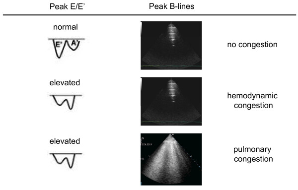
For many years the lung has been considered off-limits for ultrasound. However, it has been recently shown that lung ultrasound (LUS) may represent a useful tool for the evaluation of many pulmonary conditions in cardiovascular disease. The main application of LUS for the cardiologist is the assessment of B-lines. B-lines are reverberation artifacts, originating from water-thickened pulmonary interlobular septa. Multiple B-lines are present in pulmonary congestion, and may help in the detection, semiquantification and monitoring of extravascular lung water, in the differential diagnosis of dyspnea, and in the prognostic stratification of chronic heart failure and acute coronary syndromes.
Figures
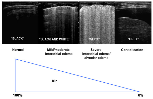
Physical basis of lung ultrasound. The less air is in the lung, the easier is the detection of lung abnormalities by ultrasound.

Methodology for lung ultrasound evaluation. Thoracic scanning areas for semiquantitative assessment of B-lines. (Modified from Jambrik et al, 2004 [7]).
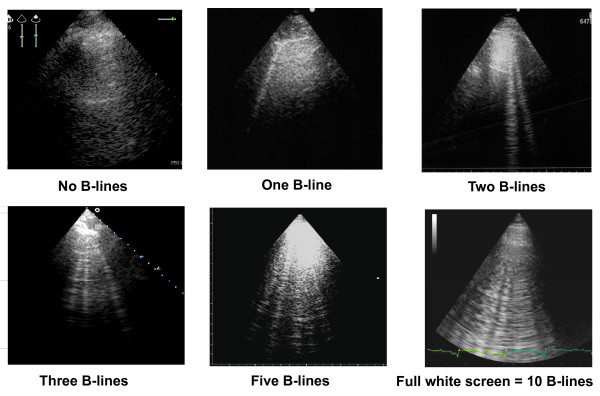
How to enumerate B-lines. Each hyperechogenic vertical stripe, spreading from the pleural line and extending to the edge of the screen, is a B-line. When using a cardiac probe, a whole white screen is considered as corresponding to a plateau value of 10 B-lines.
Alveolar-capillary membrane stress echo. The additional value of B-lines evaluation during stress echocardiography.
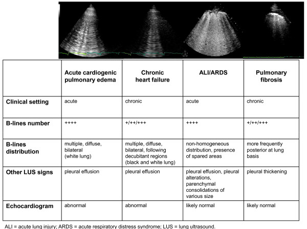
How to distinguish different etiologies of interstitial syndrome by lung ultrasound.
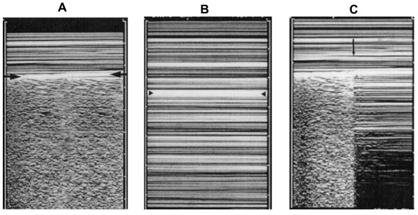
A. Normal lung pattern on M-mode: the seashore sign. The motionless superficial layers generate horizontal lines (the waves). The deep artifacts follow the lung sliding, hence the sandy pattern. B. Exclusively horizontal lines are displayed, indicating complete absence of dynamics at the level of, and below, the pleural line, a pattern called the stratosphere sign. C. M-mode evaluation of the lung point: a sudden change from the seashore to the stratosphere sign is clearly visible (arrow). (Modified from Lichtenstein et al, 2000 [48]).
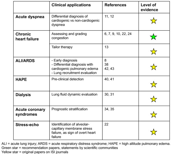
Overview of the main clinical applications of lung ultrasound for the cardiologist.
References
-
- Harrison's principles of internal medicine. 17. New York, McGraw-Hill; 2008.
-
- Lichtenstein DA. General Ultrasound in the Critically Ill. II. Berlin, Springer Verlag; 2007.
-
- Ziskin MC, Thickman DI, Goldenberg NJ, Lapayowker MS, Becker JM. The comet tail artifact. J Ultrasound Med. 1982;1:1–7. - PubMed
-
- Thickman DI, Ziskin MC, Goldenberg NJ, Linder BE. Clinical manifestations of the comet tail artifact. J Ultrasound Med. 1983;2:225–30. - PubMed
-
- Targhetta R, Chavagneux R, Balmes P, Lemerre C, Mauboussin JM, Bourgeois JM, Pourcelot L. Sonographic lung surface evaluation in pulmonary sarcoidosis: preliminary results. J Ultrasound Med. 1994;13:381–8. - PubMed
Publication types
MeSH terms
LinkOut - more resources
Full Text Sources
Other Literature Sources
Medical
