Review
. 2011 Jul;1807(7):788-802.
doi: 10.1016/j.bbabio.2011.02.006. Epub 2011 Feb 23.

The Q cycle of cytochrome bc complexes: a structure perspective

Affiliations
Review

The Q cycle of cytochrome bc complexes: a structure perspective

William A Cramer et al. Biochim Biophys Acta. 2011 Jul.

Abstract

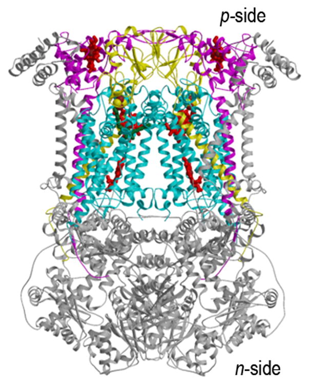
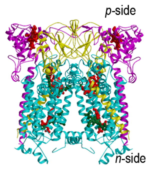
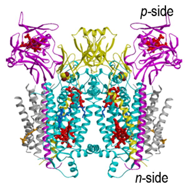
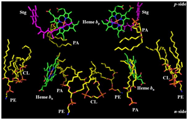
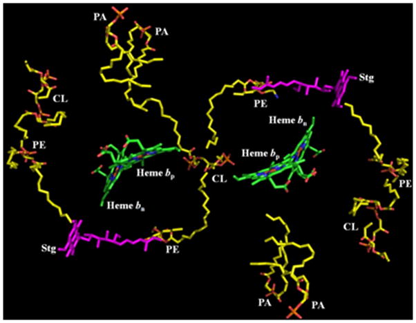
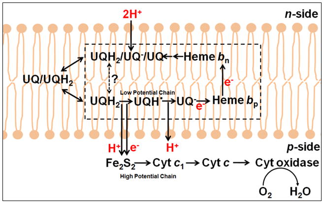
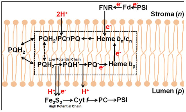
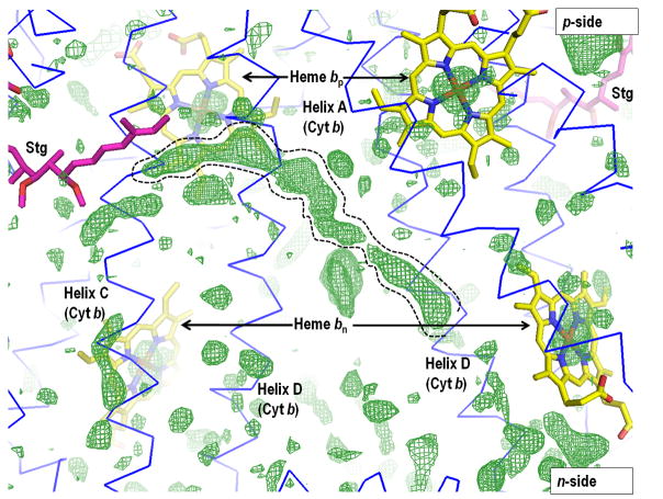
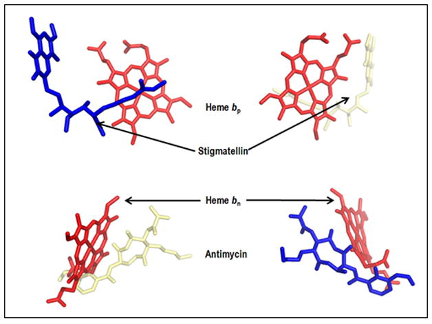
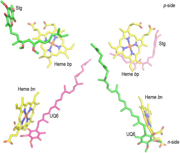
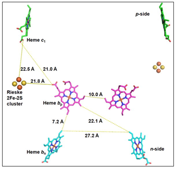
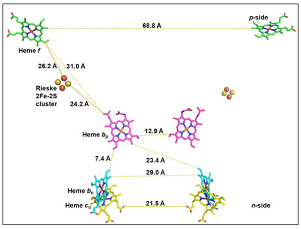
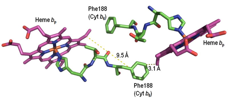
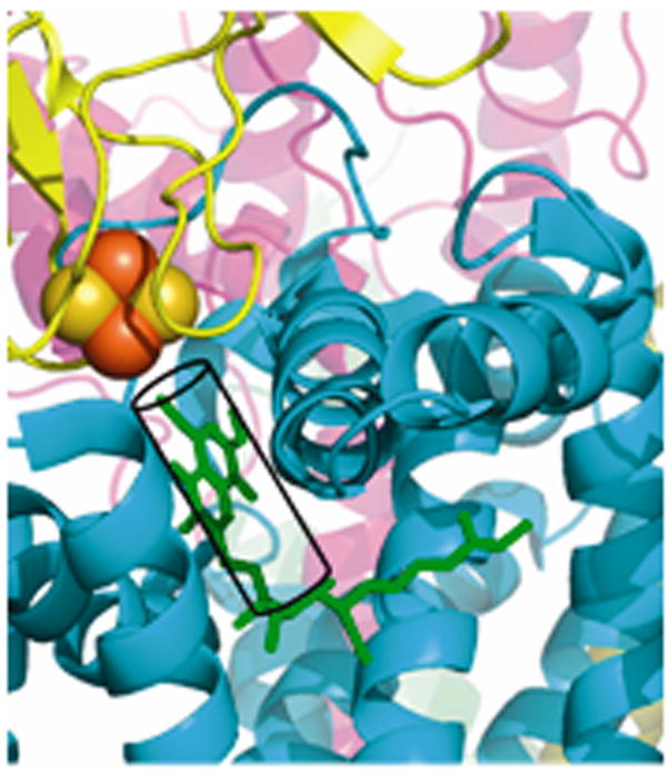
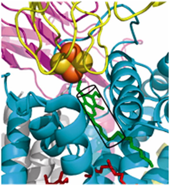
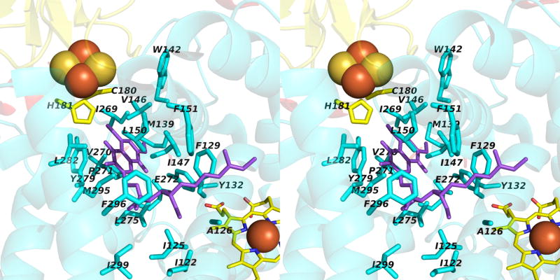
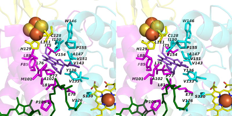
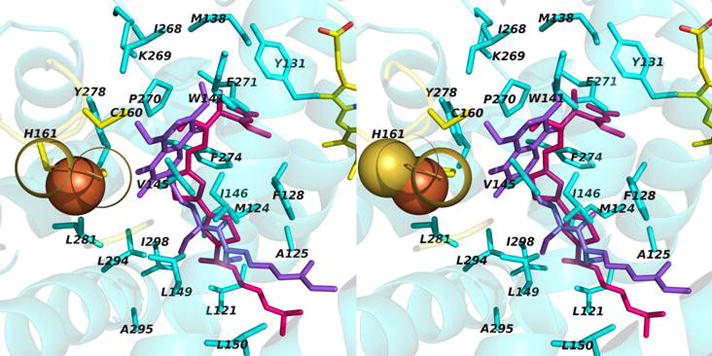
Aspects of the crystal structures of the hetero-oligomeric cytochrome bc(1) and b(6)f ("bc") complexes relevant to their electron/proton transfer function and the associated redox reactions of the lipophilic quinones are discussed. Differences between the b(6)f and bc(1) complexes are emphasized. The cytochrome bc(1) and b(6)f dimeric complexes diverge in structure from a core of subunits that coordinate redox groups consisting of two bis-histidine coordinated hemes, a heme b(n) and b(p) on the electrochemically negative (n) and positive (p) sides of the complex, the high potential [2Fe-2S] cluster and c-type heme at the p-side aqueous interface and aqueous phase, respectively, and quinone/quinol binding sites on the n- and p-sides of the complex. The bc(1) and b(6)f complexes diverge in subunit composition and structure away from this core. b(6)f Also contains additional prosthetic groups including a c-type heme c(n) on the n-side, and a chlorophyll a and β-carotene. Common structure aspects; functions of the symmetric dimer. (I) Quinone exchange with the bilayer. An inter-monomer protein-free cavity of approximately 30Å along the membrane normal×25Å (central inter-monomer distance)×15Å (depth in the center), is common to both bc(1) and b(6)f complexes, providing a niche in which the lipophilic quinone/quinol (Q/QH(2)) can be exchanged with the membrane bilayer. (II) Electron transfer. The dimeric structure and the proximity of the two hemes b(p) on the electrochemically positive side of the complex in the two monomer units allow the possibility of two alternate routes of electron transfer across the complex from heme b(p) to b(n): intra-monomer and inter-monomer involving electron cross-over between the two hemes b(p). A structure-based summary of inter-heme distances in seven bc complexes, representing mitochondrial, chromatophore, cyanobacterial, and algal sources, indicates that, based on the distance parameter, the intra-monomer pathway would be favored kinetically. (III) Separation of quinone binding sites. A consequence of the dimer structure and the position of the Q/QH(2) binding sites is that the p-side QH(2) oxidation and n-side Q reduction sites are each well separated. Therefore, in the event of an overlap in residence time by QH(2) or Q molecules at the two oxidation or reduction sites, their spatial separation would result in minimal steric interference between extended Q or QH(2) isoprenoid chains. (IV) Trans-membrane QH(2)/Q transfer. (i) n/p-side QH(2)/Q transfer may be hindered by lipid acyl chains; (ii) the shorter less hindered inter-monomer pathway across the complex would not pass through the center of the cavity, as inferred from the n-side antimycin site on one monomer and the p-side stigmatellin site on the other residing on the same surface of the complex. (V) Narrow p-side portal for QH(2)/Q passage. The [2Fe-2S] cluster that serves as oxidant, and whose histidine ligand serves as a H(+) acceptor in the oxidation of QH(2), is connected to the inter-monomer cavity by a narrow extended portal, which is also occupied in the b(6)f complex by the 20 carbon phytyl chain of the bound chlorophyll.


Figures

Fig. 1
Fig. 1
Structures of the cytochrome bc1 complex from the electron transport chain of (A) yeast mitochondria (PDB: 3CX5(15)), and (B) the purple photosynthetic bacterium, Rb. sphaeroides, with bound antimycin and stigmatellin (2QJP, (14)); (C) native b6f complex from the cyano-bacterium, M. laminosus (2E74, (19)). The ribbon diagrams show the common central structure. Color code: (yellow) Rieske protein with cluster-containing peripheral domain on one monomer and its TMH spanning the width of the other; other colors: b6f -cyt f and bc1-cyt. c1, magenta; cyt b and b6f -subunit IV, cyan.
Fig. 1
Fig. 1
Structures of the cytochrome bc1 complex from the electron transport chain of (A) yeast mitochondria (PDB: 3CX5(15)), and (B) the purple photosynthetic bacterium, Rb. sphaeroides, with bound antimycin and stigmatellin (2QJP, (14)); (C) native b6f complex from the cyano-bacterium, M. laminosus (2E74, (19)). The ribbon diagrams show the common central structure. Color code: (yellow) Rieske protein with cluster-containing peripheral domain on one monomer and its TMH spanning the width of the other; other colors: b6f -cyt f and bc1-cyt. c1, magenta; cyt b and b6f -subunit IV, cyan.
Fig. 2
Fig. 2
Arrangement of lipids, 2 PE, 2 PA, 1.5 CL per monomer, in the yeast cytochrome bc1 complex (PDB, 3CX5; (15)); (A) side and (B) top view. 1.5 molar stoichiometry of cardiolipin (CL), determined from the 3CX5 crystal structure, is a consequence of sharing one CL at the n-side interface between the two monomers.
Fig. 2
Fig. 2
Arrangement of lipids, 2 PE, 2 PA, 1.5 CL per monomer, in the yeast cytochrome bc1 complex (PDB, 3CX5; (15)); (A) side and (B) top view. 1.5 molar stoichiometry of cardiolipin (CL), determined from the 3CX5 crystal structure, is a consequence of sharing one CL at the n-side interface between the two monomers.
Fig. 3
Fig. 3
Q cycle models for electron transfer and proton translocation through (A) the bc1 complex in the respiratory chain (176) and the purple photosynthetic bacteria,(48) (reaction sequence (Table 3A1-3) and (B) the b6f complex that functions in oxygenic photosynthesis (Table 3B). The original “Q cycle” model (172, 174) for proton translocation, formulated in the aftermath of the experiment of the discovery of oxidant-induced reduction of heme b (171), focused on the mitochondrial bc1 complex. Fundamental features of the classical Q cycle are: (i) the [2Fe-2S] complex on the p-side of the complex that functions as the one electron oxidant of the two electron lipophilic quinol electron and proton donor, resulting in a bifurcated pathway into high and low potential chains; (ii) the high potential segment of the bifurcated pathway, initiated by electron transfer to cytochrome c1 or f, which transfers one electron to the high potential electron terminal acceptor, (A) cytochrome oxidase or (B) photosystem I, while generating the semiquinone; (iii) the semiquinone donates the second electron to the two trans-membrane hemes b, bp and bn, in the low potential segment of the bifurcated chain that reduces a quinone or semiquinone (53) bound at the Qn site.
Fig. 3
Fig. 3
Q cycle models for electron transfer and proton translocation through (A) the bc1 complex in the respiratory chain (176) and the purple photosynthetic bacteria,(48) (reaction sequence (Table 3A1-3) and (B) the b6f complex that functions in oxygenic photosynthesis (Table 3B). The original “Q cycle” model (172, 174) for proton translocation, formulated in the aftermath of the experiment of the discovery of oxidant-induced reduction of heme b (171), focused on the mitochondrial bc1 complex. Fundamental features of the classical Q cycle are: (i) the [2Fe-2S] complex on the p-side of the complex that functions as the one electron oxidant of the two electron lipophilic quinol electron and proton donor, resulting in a bifurcated pathway into high and low potential chains; (ii) the high potential segment of the bifurcated pathway, initiated by electron transfer to cytochrome c1 or f, which transfers one electron to the high potential electron terminal acceptor, (A) cytochrome oxidase or (B) photosystem I, while generating the semiquinone; (iii) the semiquinone donates the second electron to the two trans-membrane hemes b, bp and bn, in the low potential segment of the bifurcated chain that reduces a quinone or semiquinone (53) bound at the Qn site.
Fig. 4
Fig. 4
(A) Presence of lipid-like molecules in the inter-monomer cavity of yeast cytochrome bc1 complex (PDB 3CX5). The outlined density may correspond to an acyl chain of a lipid or detergent molecule or it may be attributed to the isoprenoid tail of a ubiquinone molecule (as found in the Qn site of the yeast bc1 complex (PDB, 1KB9). Figure generated in PyMol from PDB 3CX5 and its Fo-Fc map contoured at + 3.0 sigma. Negative densities were not included in the analysis. (B) n- and p- side binding sites of quinone analogue inhibitors, antimycin A and stigmatellin [PDB: 1PPJ (216) or 1NTZ (10), which are on the same side (yellow or blue) of the dimeric complex, implying that if a trans-complex quinone pathway operates for electron and proton transfer, it would be inter-monomer. (C) Yeast bc1 complex (PDB 1KB9) showing (side-view) cross-over of ubiquinone isoprenoid tail (UQ-6, bound at Qn site) from one monomer across the inter-monomer cavity, to the Qp site portal in the other monomer, located by presence of quinone analog stigmatellin (Stg). The Stg and UQ-6 pair colored magenta is positioned on one face of the bc1 dimer, while that colored green lies on the other.
Fig. 4
Fig. 4
(A) Presence of lipid-like molecules in the inter-monomer cavity of yeast cytochrome bc1 complex (PDB 3CX5). The outlined density may correspond to an acyl chain of a lipid or detergent molecule or it may be attributed to the isoprenoid tail of a ubiquinone molecule (as found in the Qn site of the yeast bc1 complex (PDB, 1KB9). Figure generated in PyMol from PDB 3CX5 and its Fo-Fc map contoured at + 3.0 sigma. Negative densities were not included in the analysis. (B) n- and p- side binding sites of quinone analogue inhibitors, antimycin A and stigmatellin [PDB: 1PPJ (216) or 1NTZ (10), which are on the same side (yellow or blue) of the dimeric complex, implying that if a trans-complex quinone pathway operates for electron and proton transfer, it would be inter-monomer. (C) Yeast bc1 complex (PDB 1KB9) showing (side-view) cross-over of ubiquinone isoprenoid tail (UQ-6, bound at Qn site) from one monomer across the inter-monomer cavity, to the Qp site portal in the other monomer, located by presence of quinone analog stigmatellin (Stg). The Stg and UQ-6 pair colored magenta is positioned on one face of the bc1 dimer, while that colored green lies on the other.
Fig. 5
Fig. 5
Possible pathways for electron transfer. Intra-and inter-monomer edge-edge distances for: (A) yeast bc1 (PDB: 3CX5); (B) cyanobacterial b6f (PDB: 2E74) complex. (C, D) Center-center (Fe-Fe) connection via histidine ligands, and (C) an intra-monomerTyr184–Tyr184 bridge in yeast bc1 (PDB 3CX5), and (D) a Phe188–Phe188 bridge in the M. laminosus b6f complex.
Fig. 5
Fig. 5
Possible pathways for electron transfer. Intra-and inter-monomer edge-edge distances for: (A) yeast bc1 (PDB: 3CX5); (B) cyanobacterial b6f (PDB: 2E74) complex. (C, D) Center-center (Fe-Fe) connection via histidine ligands, and (C) an intra-monomerTyr184–Tyr184 bridge in yeast bc1 (PDB 3CX5), and (D) a Phe188–Phe188 bridge in the M. laminosus b6f complex.
Fig. 6
Fig. 6
Narrow p-side quinol/quinone binding niche to access/exit the p-side [2Fe-2S] electron/proton acceptor in cytochrome bc complexes: (A): Stigmatellin (green) in a p-side portal in cytochrome bc1 complex (PDB 3CX5); (B) Tridecyl-stigmatellin (green) in a narrow portal near the p-side of the M. laminosus cyt b6f complex (PDB, 2E76); chlorophyll a shown in red, partly occluding the portal; (C, D) Expanded views (stereo) of p-side Q/QH2 entry/exit portal showing all residues within 4 Å of stigmatellin (colored violet, as in panels A, B) in (C) the yeast bc1 (PDB 3CX5) complex, and (D) the M. laminosus b6f complex (PDB: 2E76), showing the residues around tridecyl-stigmatellin (colored violet); the chlorophyll a phytyl chain (colored green) is shown occupying a portion of the portal. (E) Overlap (stereo) of p-side stigmatellin (PDB: 1SQX) and myxathiozol (1SQP) sites in the bovine mitochondrial bc1 complex.
Fig. 6
Fig. 6
Narrow p-side quinol/quinone binding niche to access/exit the p-side [2Fe-2S] electron/proton acceptor in cytochrome bc complexes: (A): Stigmatellin (green) in a p-side portal in cytochrome bc1 complex (PDB 3CX5); (B) Tridecyl-stigmatellin (green) in a narrow portal near the p-side of the M. laminosus cyt b6f complex (PDB, 2E76); chlorophyll a shown in red, partly occluding the portal; (C, D) Expanded views (stereo) of p-side Q/QH2 entry/exit portal showing all residues within 4 Å of stigmatellin (colored violet, as in panels A, B) in (C) the yeast bc1 (PDB 3CX5) complex, and (D) the M. laminosus b6f complex (PDB: 2E76), showing the residues around tridecyl-stigmatellin (colored violet); the chlorophyll a phytyl chain (colored green) is shown occupying a portion of the portal. (E) Overlap (stereo) of p-side stigmatellin (PDB: 1SQX) and myxathiozol (1SQP) sites in the bovine mitochondrial bc1 complex.

References

    1. Berry EA, Huang L-S, Earnest TN, Jap BK. X-ray diffraction by crystals of beef heart ubiquinol:cytochrome c oxidoreductase. J Mol Biol. 1992;224:1161–1166. - PubMed
    1. Xia D, Yu CA, Deisenhofer J, Xia J-Z, Yu L. Three dimensional structure of beef-heart mitochondrial cytochrome bc1 complex. Biophys J. 1996;70:253a. - PubMed
    1. Xia D, Yu C-A, Kim H, Xia J-Z, Kachurin AM, Yu L, Deisenhofer J. Crystal structure of the cytochrome bc1 complex from bovine heart mitochondria. Science (New York, NY. 1997;277:60–66. - PubMed
    1. Iwata S, Lee JW, Okada K, Lee JK, Iwata M, Rasmussen B, Link TA, Ramaswamy S, Jap BK. Complete structure of the 11-subunit bovine mitochondrial cytochrome bc1 complex. Science (New York, NY. 1998;281:64–71. - PubMed
    1. Zhang Z, Huang L, Shulmeister VM, Chi YI, Kim KK, Hung LW, Crofts AR, Berry EA, Kim SH. Electron transfer by domain movement in cytochrome bc1. Nature. 1998;392:677–684. - PubMed

Publication types

MeSH terms

LinkOut - more resources