Review
. 2011 May;70(1):2-8.
doi: 10.1016/j.neures.2011.02.004. Epub 2011 Feb 23.

Excitation-transcription coupling in sympathetic neurons and the molecular mechanism of its initiation

Affiliations
Review

Excitation-transcription coupling in sympathetic neurons and the molecular mechanism of its initiation

Huan Ma et al. Neurosci Res. 2011 May.

Abstract

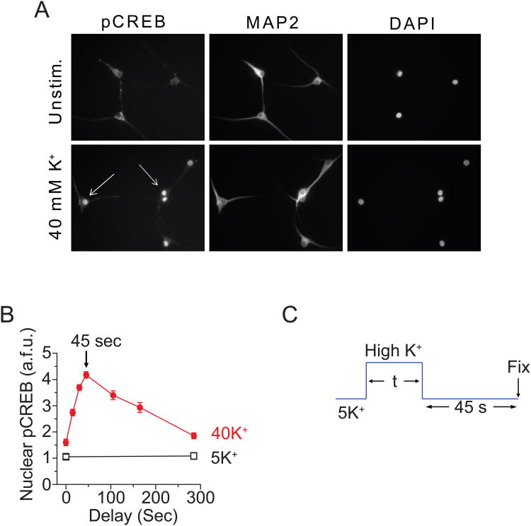
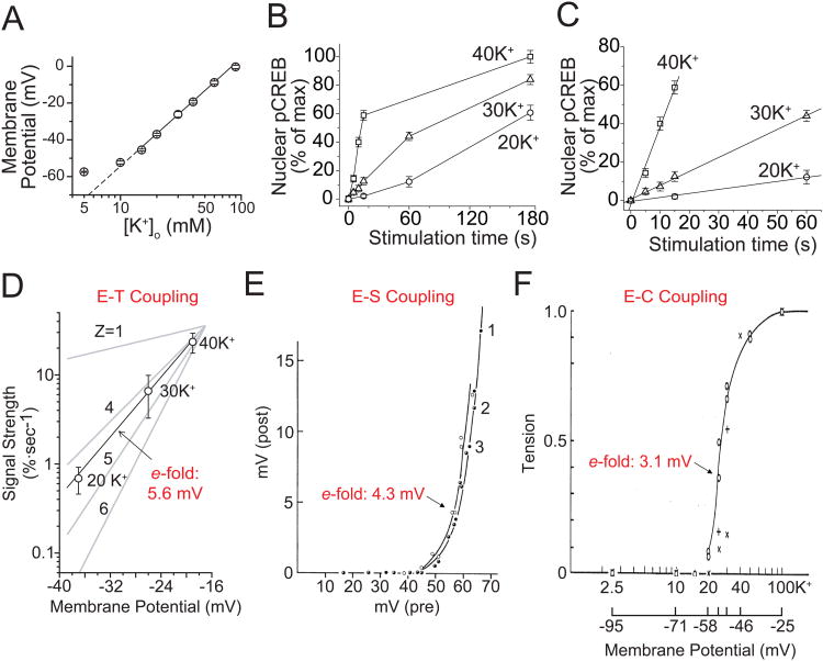
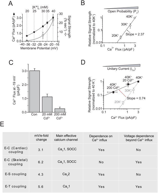
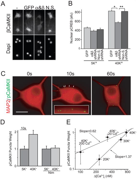
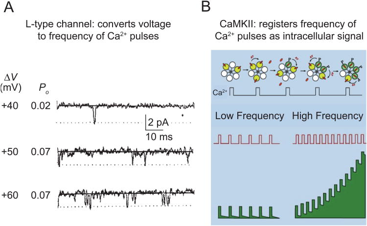
In excitable cells, membrane depolarization and activation of voltage-gated Ca²+ (Ca(V)) channels trigger numerous cellular responses, including muscle contraction, secretion, and gene expression. Yet, while the mechanisms underlying excitation-contraction and excitation-secretion coupling have been extensively characterized, how neuronal activity is coupled to gene expression has remained more elusive. In this article, we will discuss recent progress toward understanding the relationship between patterns of channel activity driven by membrane depolarization and activation of the nuclear transcription factor CREB. We show that signaling strength is steeply dependent on membrane depolarization and is more sensitive to the open probability of Ca(V) channels than the Ca²+ entry itself. Furthermore, our data indicate that by decoding Ca(V) channel activity, CaMKII (a Ca²+/calmodulin-dependent protein kinase) links membrane excitation to activation of CREB in the nucleus. Together, these results revealed some interesting and unexpected similarities between excitation-transcription coupling and other forms of excitation-response coupling.


Figures

Fig. 1
Fig. 1. Quantification of pCREB signaling with a time delay
(A) SCG neurons (cultured 5 days) were either mock-stimulated in 5 mM K+ Tyrode solution (containing 2 mM Ca2+) or stimulated with solutions containing 40 mM K+ for 180 s. After stimulation, cells were immediately fixed and stained for pCREB and MAP2; nuclei were counterstained with DAPI. Arrows indicate strongly activated neurons. (B) Neurons were stimulated with 5 or 40 mM K+ for 10 s followed by a variable time delay before fixation. Peak pCREB levels occurred 45 s after stimulation and decayed thereafter, becoming nearly indistinguishable from baseline within 5 min. (C) To ensure that the pCREB level measured after a given stimulation was a maximal response, we stimulated the cells for different time with a fixed 45 s delay in 5 mM K+ Tyrode solution. (Modified from Wheeler et al., 2008 or Wheeler et al. unpublished data.)
Fig. 2
Fig. 2. Strength of signaling to CREB in SCG neurons
(A) Membrane potential plotted against [K+]o. The slope of the linear fit is 58.1 mV per 10-fold change in [K+]o, consistent with the Nernst equation. (B) Mean pCREB levels plotted against stimulation time (with a 45 s delay as determined in Figure 1); [K+]o as indicated. (C) Linear fits of initial data points, using the slope as an index of CREB signal strength, pCREB. (D) CREB signal strength plotted against membrane voltage. 100% is the full change in pCREB from baseline to maximal levels. A linear fit shows that signal strength is steeply voltage dependent, as if governed by a gating particle with a valence (z) between 4 and 5 (corresponding to 5.6 mV/e-fold change in signal strength). (E) E-S coupling is also sharply dependent on voltage, with an e-fold change per 4.3 mV. Curves 1 (full circles), curves 2 (half-filled circles) and 3 (open circles) were obtained with pre-spikes, local pulses of 1 msec and 2 msec duration, respectively. (Katz et al., 1967). (F) The e-fold change for E-C coupling is per 3.1 mV (Hodgkin et al., 1960). (A-D Modified from Wheeler et al., 2008; E modified from Katz et al., 1967; F modified from Hodgkin et al., 1960.)
Fig. 3
Fig. 3. CREB signal strength is steeply dependent on channel Po but is largely independent of unitary Ca2+flux
(A) Whole-cell Ca2+ flux (measured with 2 mM Ca2+ and normalized to cell capacitance) and CREB signal strength plotted versus membrane potential. Solid lines are single exponential fits. Dashed lines indicate the corresponding [K+]o yielding the given membrane potentials, derived from Figure 2A. (B) Log-log plot of signal strength versus Ca2+ flux for the [K+]o indicated. ]The solid line is a linear fit of the data. (Almers) Whole-cell Ca2+ flux, in the presence of 0, 20, or 200 μM Cd2+ (Almers)CREB signal strength for 40 K+ with 0, 20, or 200 μM Cd2+. The solid line is a linear fit of the Cd2+ data. For comparison, the dashed line and gray data points are reproduced from B. (E) Some salient features of the three major forms of excitation-response coupling. SOCC, store-operated calcium channel. (Modified from Wheeler et al., 2008.)
Fig. 4
Fig. 4. CaMKII is critical for CREB signaling
(A) SCG neurons infected with lentiviruses expressing GFP alone, shRNAs against α and βCaMKII, or nonsilencing control shRNAs, and stained with an antibody against βCaMKII; nuclei were counterstained with DAPI. (B) pCREB levels from neurons stimulated for 2.5 s with 5 mM K+ or 40 mM K+ and transferred to a 5 mM K+ solution for 45 s before fixation. *p<5×10-17; **p< 5×10⁻¹⁰. (C) SCG neurons stimulated with 40 mM K+ for 0, 10, or 60 s and immediately fixed and stained for MAP2 (red) and pCaMKII (green). Arrows point to pCaMKII puncta (magnified in the inset). Background pCaMKII staining was subtracted to highlight punctuate staining. Scale bar, 20 μm. (D) Nimodipine blocks pCaMKII puncta formation. (E) Plot of pCaMKII puncta weight versus the rise in bulk Ca2+ measured using Fura-2 ratiometric Ca2+ imaging for the stimulation conditions indicated. (Modified from Wheeler et al., 2008.)
Fig. 5
Fig. 5. CaM/CaMKII decodes the digital frequency code generated by CaV1 channels
(A) The frequency of CaV1 channel opening (Po) is steeply voltage-dependent (Reuter et al., 1982). (B) The activity of CaMKII increases in a steeply frequency-dependent way (top) (De Koninck and Schulman, 1998). Quantitative modeling suggests that the frequency-dependence holds for brief pulses of Ca2+ of the kind produced by the activity of CaV1 channels (Dupont et al., 2003). Thus, the combination of CaV1 and CaMKII supports a highly sensitive transduction of cell membrane potential to intracellular biochemical signaling (bottom). (A modified from Reuter et al., 1982; B was kindly provided by A. Hudmon.)

References

    1. Almers W. Gating currents and charge movements in excitable membranes. Rev Physiol Biochem Pharmacol. 1978;82:96–190. - PubMed
    1. Armstrong CM, Bezanilla FM, Horowicz P. Twitches in the presence of ethylene glycol bis(-aminoethyl ether)-N,N'-tetracetic acid. Biochimica et biophysica acta. 1972;267:605–608. - PubMed
    1. Augustine GJ. How does calcium trigger neurotransmitter release? Current opinion in neurobiology. 2001;11:320–326. - PubMed
    1. Augustine GJ, Charlton MP, Smith SJ. Calcium entry and transmitter release at voltage-clamped nerve terminals of squid. The Journal of physiology. 1985;367:163–181. - PMC - PubMed
    1. Beam KG, Franzini-Armstrong C. Functional and structural approaches to the study of excitation-contraction coupling. Methods Cell Biol. 1997;52:283–306. - PubMed

Publication types

Substances

LinkOut - more resources