. 2011 Oct;25(7):1299-304.
doi: 10.1016/j.bbi.2011.02.009. Epub 2011 Feb 23.

Epigenetic impact of simulated maternal grooming on estrogen receptor alpha within the developing amygdala

Affiliations

Epigenetic impact of simulated maternal grooming on estrogen receptor alpha within the developing amygdala

Michelle N Edelmann et al. Brain Behav Immun. 2011 Oct.

Abstract

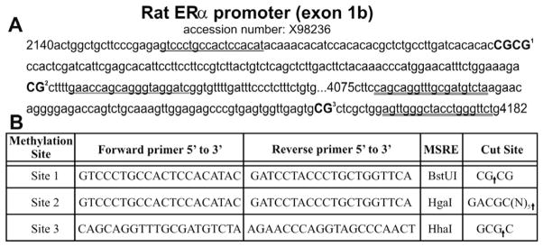
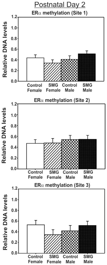
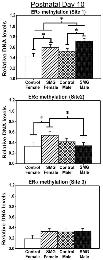
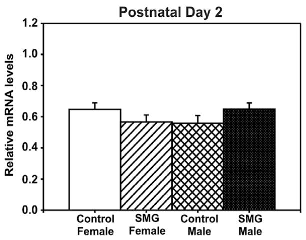
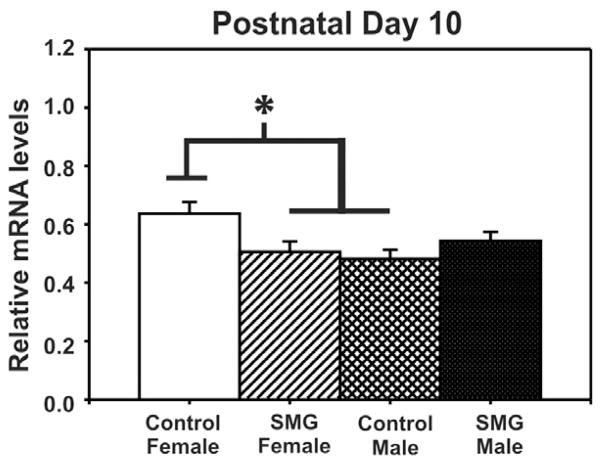
Variations in maternal care alter the developmental programming of some genes by creating lasting differences in DNA methylation patterns, such as the estrogen receptor alpha (ERα) promoter region. Interestingly, mother rats preferentially lick and groom their male offspring more than females; therefore, we questioned whether the somatosensory stimuli associated with maternal grooming influences potential sex differences in DNA methylation patterns within the developing amygdala, an area important for socioemotional processing. We report a sex difference in the DNA methylation pattern of specific CpG sites of the ERα promoter region within the developing amygdala. Specifically, males have higher levels of ERα promoter methylation contrasted to females. Increasing the levels of maternal stimuli in females masculinized ERα promoter methylation patterns to male-like levels. As expected, higher levels of ERα promoter methylation were associated with lower ERα mRNA levels. These data provide further evidence that the early neonatal environment, particularly maternal care, contributes to sex differences and early programming of the neonatal brain via an epigenetic mechanism.


Figures

Fig. 1
Fig. 1
Methodological details for CpG sites analyzed within the exon 1b ERα promoter. (A) Sequence for the portion of the exon 1b ERα promoter region (GenBank Accession No. X98236) examined. Primer annealing sequences used for examining methylation at site 1 and site 2 is underlined with a single line. Primer annealing sequences used for examining methylation at site 3 is double underlined. Each methylation site examined is bolded and numbered. (B) Forward and reverse primer sequences are shown that were used to investigate the methylation status of the three sites using a methyl-sensitive restriction enzyme (MSRE). The same primers were used for examining methylation at site 1 or site 2. The nucleotide sequence cut site for each MSRE is indicated by an arrow. Methylation at these CpG sites protects against enzyme digestion and allows for PCR amplification.
Fig. 2
Fig. 2
ERα promoter methylation is not altered by simulated maternal grooming or sex on P2. No significant differences in CpG methylation were found at either of the CpG sites examined (p > 0.05).
Fig. 3
Fig. 3
ERα promoter methylation is altered by simulated maternal grooming and sex on P10. At site1, there is a sex difference in methylation where males have more methylation than females (*p < 0.05). Additionally, SMG-treated females have significantly more methylation than control-handled females (*p < 0.05) and are not statistically different than males. Interestingly, SMG also altered DNA methylation in males. At site 2, SMG-treated females had significantly more methylation than SMG-treated males (*p < 0.05). A strong trend towards SMG females having more methylation than control females was observed (#p = 0.055). There were no significant differences found at the third CpG site (p > 0.05).
Fig. 4
Fig. 4
ERα mRNA levels are not altered by simulated maternal grooming or sex on P2. No significant difference in ERα mRNA levels was observed (p > 0.05).
Fig. 5
Fig. 5
ERα mRNA levels are altered by simulated maternal grooming and sex on P10. Control females expressed more ERα mRNA than SMG-treated females (*p < 0.05) or control males (*p < 0.01). There is no statistical difference between SMG-treated females and males.

Similar articles

Cited by

References

    1. Amateau SK, Alt JJ, Stamps CL, McCarthy MM. Brain estradiol content in newborn rats: sex differences, regional heterogeneity, and possible de novo synthesis by the female telencephalon. Endocrinology. 2004;145:2906–2917. - PubMed
    1. Auger AP, Jessen HM, Edelmann MN. Epigenetic organization of brain sex differences and juvenile social play behavior. Horm Behav. 2010 doi: 10.1016/j.yhbeh.2010.06.017. [Epub ahead of print] - DOI - PMC - PubMed
    1. Auger AP, Olesen KM. Brain sex differences and the organisation of juvenile social play behaviour. J Neuroendocrinol. 2009;21:519–525. - PMC - PubMed
    1. Barclay JL, Nelson CN, Ishikawa M, Murray LA, Kerr LM, McPhee TR, Powell EE, Waters MJ. GH-dependent STAT5 signaling plays an important role in hepatic lipid metabolism. Endocrinology. 2011;152:181–192. - PubMed
    1. Bonefeld BE, Elfving B, Wegener G. Reference genes for normalization: a study of rat brain tissue. Synapse. 2008;62:302–309. - PubMed

Publication types

Substances