

doi: 10.1016/j.vaccine.2011.02.023.
Epub 2011 Feb 23.
Deletion of the A35 gene from Modified Vaccinia Virus Ankara increases immunogenicity and isotype switching
Affiliations
- PMID: 21352940
- PMCID: PMC3078999
- DOI: 10.1016/j.vaccine.2011.02.023
Item in Clipboard
Deletion of the A35 gene from Modified Vaccinia Virus Ankara increases immunogenicity and isotype switching
Vaccine.
.
Abstract
We show here that the immunogenicity of the Modified Vaccinia Ankara MVA vaccine strain can be improved by deletion of the A35 gene, without diminishing the ability of the virus to replicate. Deletion of the A35 gene resulted in increased virus-specific immunoglobulin production, class switching to IgG isotypes, and virus-specific IFNγ-secreting splenocytes. The MVA35 deletion virus provided excellent protective efficacy against virulent virus challenge. These results suggest that A35 deletion mutant strains will have superior vaccine performance for poxvirus vaccines as well as platform vaccines for other infectious diseases and cancer.
Copyright © 2011 Elsevier Ltd. All rights reserved.
Figures
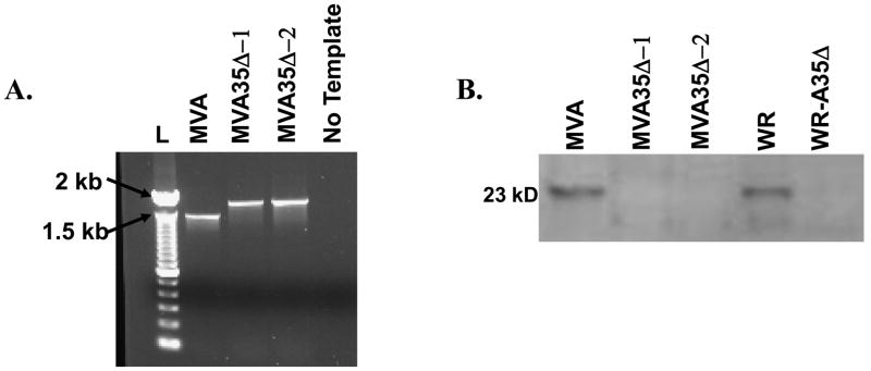
a) PCR. MVA-infected cells were transfected with a recombinant PCR fragment containing the E. coli gpt gene inserted between the A35 flanking regions and recombinant viruses were selected in mycophenolic acid-containing media. Virus crude stocks were PCR analyzed using primers in the A35 flanking regions. Wild type A35 locus yields a product of 1400 kbp size and the mutants with gpt inserted yield a size of approx 1900 kbp. L=ladder, b) Western blot showing that A35 is not expressed in MVA35Δ-infected cells. BHK-21 cells were infected with listed viruses at an MOI of 20 for 2 h and analyzed by SDS-PAGE. Blots were incubated with rabbit anti-A35 antibody at a 1:1000 dilution and developed with an anti-rabbit alkaline phosphatase-conjugated secondary antibody.
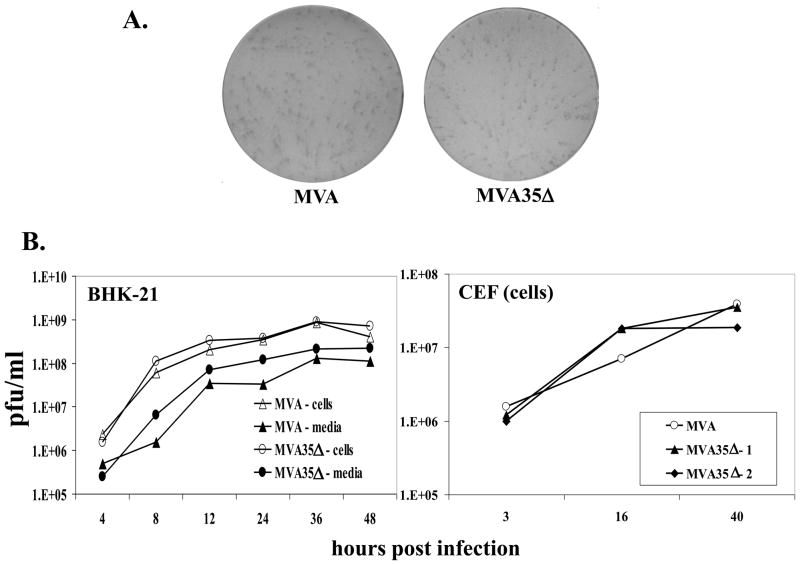
a) MVA and MVA35Δ mutant viruses form similarly sized foci on BHK-21 monolayers. Virus-infected cells were visualized by immunostaining. b) One step growth curve. BHK-21 cells or CEF were infected (MOI=10) with MVA, MVA35Δ–1, or MVA35Δ-2, and the amount of virus associated with cells and in the supernatants was titered on BHK-21 cells at various times post infection.
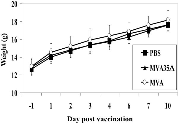
Groups of mice (n=5) were infected i.m. with 107 pfu/mouse of MVA or MVA35Δ virus, or mock-vaccinated with PBS and weighed (g + SD) for 4 weeks. No mice lost weight or showed signs of illness.
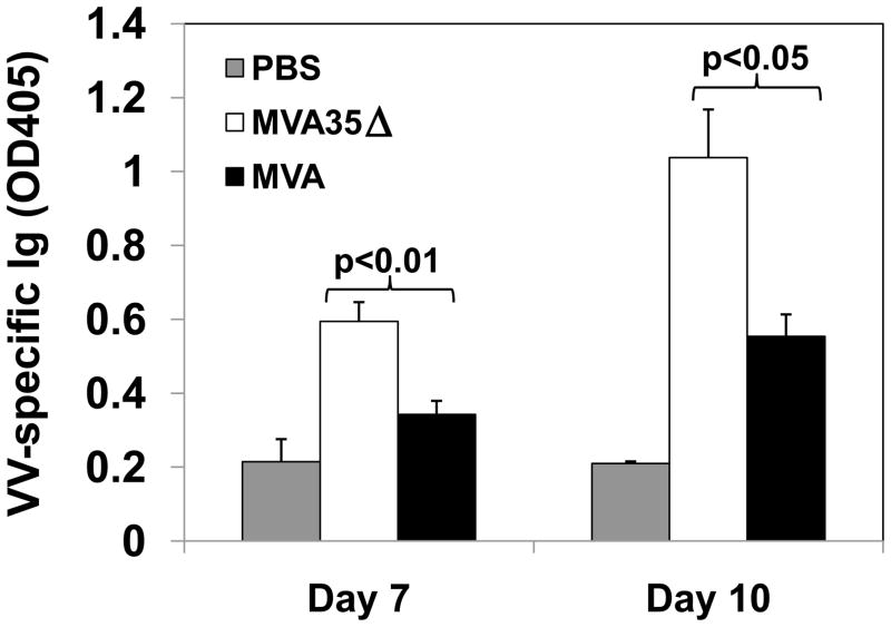
Mice (PBS n=3 day 7, n=4 day 10; MVA35Δ n=4; MVA n=4) were infected i.m. with MVA or MVA35Δ virus, and blood was collected via cardiac stick on various days pi. Sera were titrated 1:2, dilutions ranging from 1:20–1:2560. Total VACV-Ig was measured by ELISA on VACV coated plates for day 7 and 10. Data show the average absorbance (day 7 – 1:80, day 10 – 1:20) at 405 nm (+/− SEM).
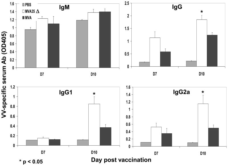
Mice (n=3–5) were infected i.m. with MVA or MVA35Δ virus, and blood was collected on various days pi. Sera were diluted 1:2, resulting in dilutions ranging from 1:10–1:1280. VACV-specific IgM, IgG, IgG1, and IgG2a were measured by ELISA on VACV coated plates for day 7 and 10. Data show the average absorbance (1:20 dilution) at 405 nm (+/− SEM).
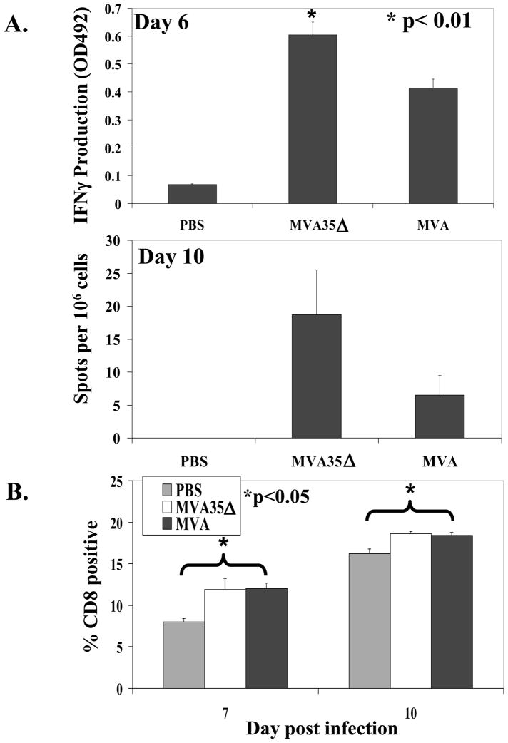
a) IFNγ production. On days 6 and 10 pi (i.m.), the spleens from 5 mice/group were harvested and splenocytes were analyzed by ELISPOT for virus-specific IFNγ production 48 h after stimulation with VACV-WR virus. b) CD8+ cells. On days 7 and 10 pi, spleens from vaccinated mice were analyzed by flow cytometry for the percentage of CD8+ T cells. Data show the average (+/− SEM).
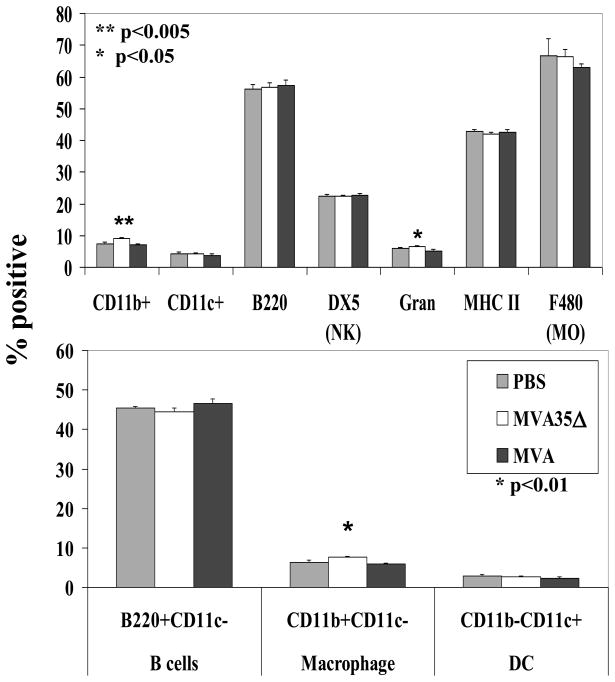
On day 6 pi, spleens from MVA and MVA35Δ-infected mice (n=5) were stained for various cell surface markers to enumerate percentage of different cell types. Data show average percentage (+/− SEM). Gran, granulocytes; NK, natural killer; MO, macrophage; DC, dendritic cell.
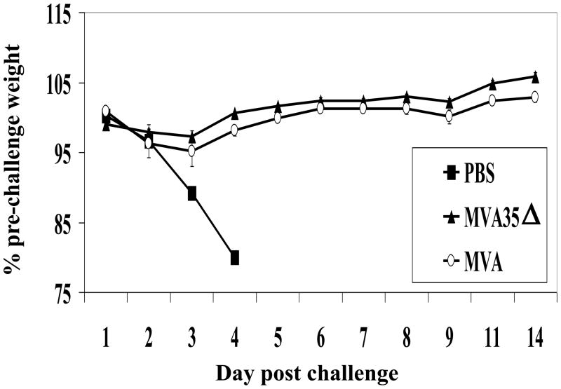
Mice (n=5) were vaccinated i.m. on day 0 with 107 pfu of MVA or MVA35Δ virus, or mock-vaccinated with PBS. Four weeks later, the mice were challenged i.n. with LD50 × 500 virulent WR virus. Data show average percent change in pre-challenge weight (+/− SEM). All mice died in the PBS mock vaccinated group.
Similar articles
-
Vaccination of BALB/c mice with Escherichia coli-expressed vaccinia virus proteins A27L, B5R, and D8L protects mice from lethal vaccinia virus challenge.J Virol. 2008 Apr;82(7):3517-29. doi: 10.1128/JVI.01854-07. Epub 2008 Jan 16. J Virol. 2008. PMID: 18199639 Free PMC article.
-
Development of smallpox vaccine candidates with integrated interleukin-15 that demonstrate superior immunogenicity, efficacy, and safety in mice.J Virol. 2007 Aug;81(16):8774-83. doi: 10.1128/JVI.00538-07. Epub 2007 Jun 6. J Virol. 2007. PMID: 17553867 Free PMC article.
-
E3L and F1L Gene Functions Modulate the Protective Capacity of Modified Vaccinia Virus Ankara Immunization in Murine Model of Human Smallpox.Viruses. 2018 Jan 4;10(1):21. doi: 10.3390/v10010021. Viruses. 2018. PMID: 29300297 Free PMC article.
-
Clinical development of Modified Vaccinia virus Ankara vaccines.Vaccine. 2013 Sep 6;31(39):4241-6. doi: 10.1016/j.vaccine.2013.03.020. Epub 2013 Mar 21. Vaccine. 2013. PMID: 23523410 Review.
-
Modified Vaccinia Virus Ankara: History, Value in Basic Research, and Current Perspectives for Vaccine Development.Adv Virus Res. 2017;97:187-243. doi: 10.1016/bs.aivir.2016.07.001. Epub 2016 Aug 1. Adv Virus Res. 2017. PMID: 28057259 Free PMC article. Review.
Cited by
-
Increased attenuation but decreased immunogenicity by deletion of multiple vaccinia virus immunomodulators.Vaccine. 2016 Sep 14;34(40):4827-34. doi: 10.1016/j.vaccine.2016.08.002. Epub 2016 Aug 17. Vaccine. 2016. PMID: 27544585 Free PMC article.
-
Orthopoxvirus genes that mediate disease virulence and host tropism.Adv Virol. 2012;2012:524743. doi: 10.1155/2012/524743. Epub 2012 Jul 30. Adv Virol. 2012. PMID: 22899927 Free PMC article.
-
Deletion of Fifteen Open Reading Frames from Modified Vaccinia Virus Ankara Fails to Improve Immunogenicity.PLoS One. 2015 Jun 8;10(6):e0128626. doi: 10.1371/journal.pone.0128626. eCollection 2015. PLoS One. 2015. PMID: 26053118 Free PMC article.
-
Waning of SARS-CoV-2 Seropositivity among Healthy Young Adults over Seven Months.Vaccines (Basel). 2022 Sep 15;10(9):1532. doi: 10.3390/vaccines10091532. Vaccines (Basel). 2022. PMID: 36146610 Free PMC article.
-
Genes that Control Vaccinia Virus Immunogenicity.Acta Naturae. 2020 Jan-Mar;12(1):33-41. doi: 10.32607/actanaturae.10935. Acta Naturae. 2020. PMID: 32477596 Free PMC article.
References
-
- Mahalingam S, Damon IK, Lidbury BA. 25 years since the eradication of smallpox: why poxvirus research is still relevant. Trends Immunol. 2004 Dec;25(12):636–9. - PubMed
-
- Lederman E, Miramontes R, Openshaw J, Olson VA, Karem KL, Marcinak J, et al. Eczema vaccinatum resulting from the transmission of vaccinia virus from a smallpox vaccinee: An investigation of potential fomites in the home environment. Vaccine. 2009 Jan 14;27(3):375–7. - PubMed
-
- Eckart RE, Love SS, Atwood JE, Arness MK, Cassimatis DC, Campbell CL, et al. Incidence and follow-up of inflammatory cardiac complications after smallpox vaccination. J Am Coll Cardiol. 2004 Jul 7;44(1):201–5. - PubMed
-
- Kemper A, Davis M, Freed G. Expected Adverse Events in a Mass Smallpox Vaccination Campaign. Effective Clinical Practice. 2002 March - PubMed
-
- Upfal MJ, Cinti S. Smallpox vaccination and adverse cardiac events. Emerg Infect Dis. 2004 May;10(5):961–2. discussion 2. - PubMed
Publication types
MeSH terms
Substances
Grants and funding
LinkOut - more resources
Full Text Sources
Medical
