Case Reports
. 2011 Mar 11;88(3):362-71.
doi: 10.1016/j.ajhg.2011.01.015. Epub 2011 Feb 25.

Exome sequencing identifies truncating mutations in human SERPINF1 in autosomal-recessive osteogenesis imperfecta

Affiliations
Case Reports

Exome sequencing identifies truncating mutations in human SERPINF1 in autosomal-recessive osteogenesis imperfecta

Jutta Becker et al. Am J Hum Genet. .

Abstract

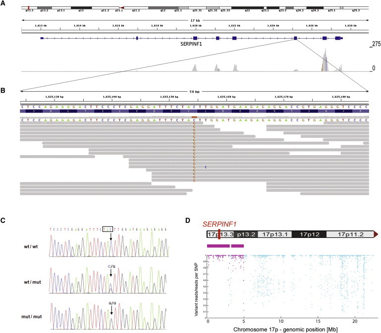
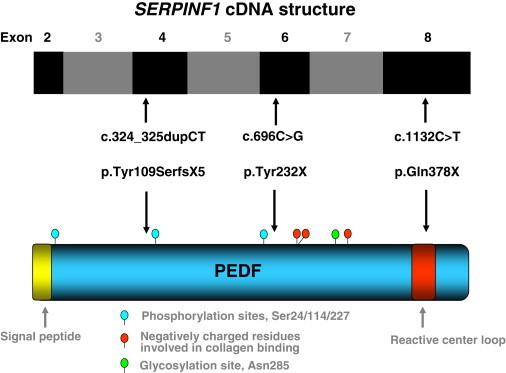
Osteogenesis imperfecta (OI) is a heterogeneous genetic disorder characterized by bone fragility and susceptibility to fractures after minimal trauma. After mutations in all known OI genes had been excluded by Sanger sequencing, we applied next-generation sequencing to analyze the exome of a single individual who has a severe form of the disease and whose parents are second cousins. A total of 26,922 variations from the human reference genome sequence were subjected to several filtering steps. In addition, we extracted the genotypes of all dbSNP130-annotated SNPs from the exome sequencing data and used these 299,494 genotypes as markers for the genome-wide identification of homozygous regions. A single homozygous truncating mutation, affecting SERPINF1 on chromosome 17p13.3, that was embedded into a homozygous stretch of 2.99 Mb remained. The mutation was also homozygous in the affected brother of the index patient. Subsequently, we identified homozygosity for two different truncating SERPINF1 mutations in two unrelated patients with OI and parental consanguinity. All four individuals with SERPINF1 mutations have severe OI. Fractures of long bones and severe vertebral compression fractures with resulting deformities were observed as early as the first year of life in these individuals. Collagen analyses with cultured dermal fibroblasts displayed no evidence for impaired collagen folding, posttranslational modification, or secretion. SERPINF1 encodes pigment epithelium-derived factor (PEDF), a secreted glycoprotein of the serpin superfamily. PEDF is a multifunctional protein and one of the strongest inhibitors of angiogenesis currently known in humans. Our data provide genetic evidence for PEDF involvement in human bone homeostasis.


Figures

Figure 1
Figure 1
Identification of a Homozygous Mutation in SERPINF1 by Exome Sequencing (A) Overview of SERPINF1 coverage by exome sequencing. Upper: An ideogram illustrating the SERPINF1 location in the chromosomal band 17p13.3 (red bar). Middle: The genomic structure and localization of SERPINF1 in more detail. Lower: The per-base coverage, with an axis to the right ranging from 0 to 275-fold coverage. (B) Detailed view of individual sequencing reads overlapping the stop mutation in exon 6. All 18 reads at the genomic position g.1,625,154 (hg18) display the homozygous stop mutation c.696C>G. (C) Validation of the SERPINF1 mutation in exon 6 by Sanger sequencing of genomic DNA. Electropherograms of a control individual, of the index patient, and of his father are shown. The position of the mutation is marked with an arrow. The affected codon is framed in black. The control individual carries the wild-type sequence (wt) in both copies of the gene. The patient's father is a heterozygous carrier of the mutation (genotype wt/mut). The patient is homozygous for the c.696C>G mutation (mut/mut). (D) Upper: Contains an ideogram of the p-arm of chromosome 17 illustrating the localization of SERPINF1. Lower : The homozygous regions on chromosome 17 at the genomic positions g.97,058-3,091,589 and g.3,299,188-4,983,922 are recognizable. Each dot represents one of the SNP markers that were included in the analysis. SNPs within a region that was called as homozygous are colored in purple. The position along the y axis indicates the ratio of the number of reads with the nonreference allele to the total number of reads; the value is 1.0 if only reference or nonreference reads are present at a given SNP position (y axis, ratio of variant reads/all reads per SNP position; x axis, genomic position along the p-arm of chromosome 17).
Figure 2
Figure 2
SERPINF1 and PEDF Scheme Schematic representation of pigment epithelium-derived factor (PEDF, NP_002606) and of the coding exons of the SERPINF1 cDNA (NM_002615.4). The locations of the exons are aligned relative to the regions of the PEDF each exon encodes. The positions of the three truncating mutations identified in OI patients are indicated by arrows. Several important protein domains described in the literature are depicted.
Figure 3
Figure 3
Radiological Features of Patient 3 (A and B) Thorax and pelvis of patient 3 at the age of 6 months showing no signs of rip or clavicular fractures, only a slightly decreased vertebral height, a small pelvis and moderately delayed development of the hips. (C and D) Radiographs of the right and left leg when the patient was 1.6 years old show severe bowing of both femurs, multiple old and new fractures, and a pseudarthrosis in the right femur. In the epiphysis, lines resulting from intravenous bisphosphonate treatment are visible. The tibiae display no severe deformities.
Figure 4
Figure 4
Radiological Features of Patient 4 (A) Hand radiogram at the age of 6 years shows no signs of fractures or skeletal dysplasia and only a mild delay of bone age. (B) The right arm at the age of 5.4 years shows severe deformities of the humerus with multiple old fractures and callus formation in the diaphysis. (C) Left hemithorax at the age of 4.3 years displays no deformities or fractures of rips. (D) Right leg at the age of 2.9 years after surgical rodding with an intramedullar telescopic rod because of a fracture. (E) Fractured right leg 3 months earlier. Also visible are the epiphyseal lines resulting from cyclic intravenous bisphosphonate treatment. (F) Left leg with a fracture after minimal trauma at the age of 2.2 years. (G) Left leg a few weeks later after surgical treatment with an intramedullar telescopic rod.

References

    1. Rauch F., Glorieux F.H. Osteogenesis imperfecta. Lancet. 2004;363:1377–1385. - PubMed
    1. Basel D., Steiner R.D. Osteogenesis imperfecta: Recent findings shed new light on this once well-understood condition. Genet. Med. 2009;11:375–385. - PubMed
    1. Barsh G.S., Byers P.H. Reduced secretion of structurally abnormal type I procollagen in a form of osteogenesis imperfecta. Proc. Natl. Acad. Sci. USA. 1981;78:5142–5146. - PMC - PubMed
    1. Byers P.H., Tsipouras P., Bonadio J.F., Starman B.J., Schwartz R.C. Perinatal lethal osteogenesis imperfecta (OI type II): A biochemically heterogeneous disorder usually due to new mutations in the genes for type I collagen. Am. J. Hum. Genet. 1988;42:237–248. - PMC - PubMed
    1. Chu M.L., Williams C.J., Pepe G., Hirsch J.L., Prockop D.J., Ramirez F. Internal deletion in a collagen gene in a perinatal lethal form of osteogenesis imperfecta. Nature. 1983;304:78–80. - PubMed

Publication types