. 2011 Mar 8;21(5):391-8.
doi: 10.1016/j.cub.2010.12.039. Epub 2011 Feb 25.

Direct binding of Cenp-C to the Mis12 complex joins the inner and outer kinetochore

Affiliations

Direct binding of Cenp-C to the Mis12 complex joins the inner and outer kinetochore

Emanuela Screpanti et al. Curr Biol. .

Abstract

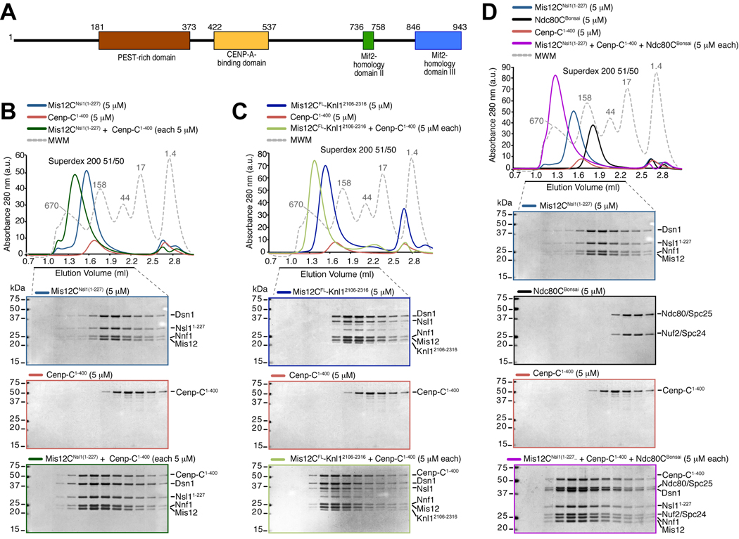
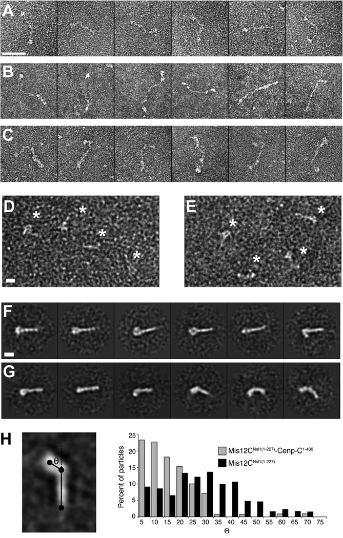
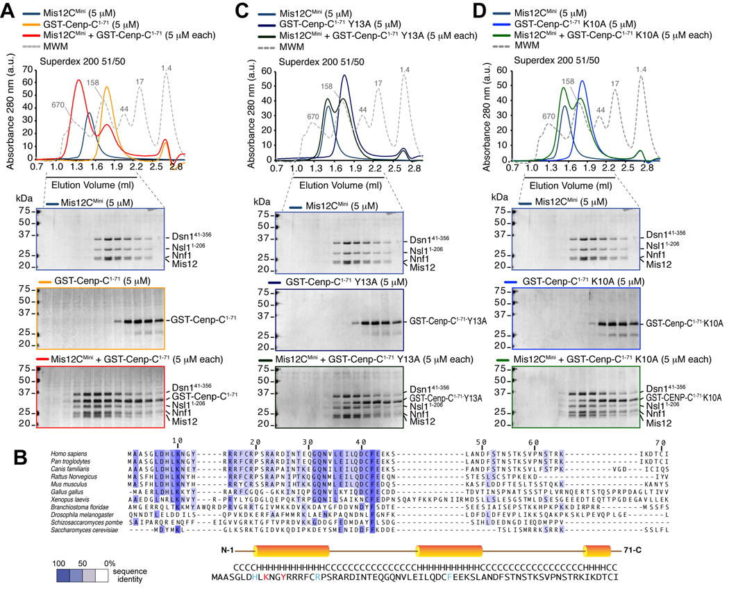
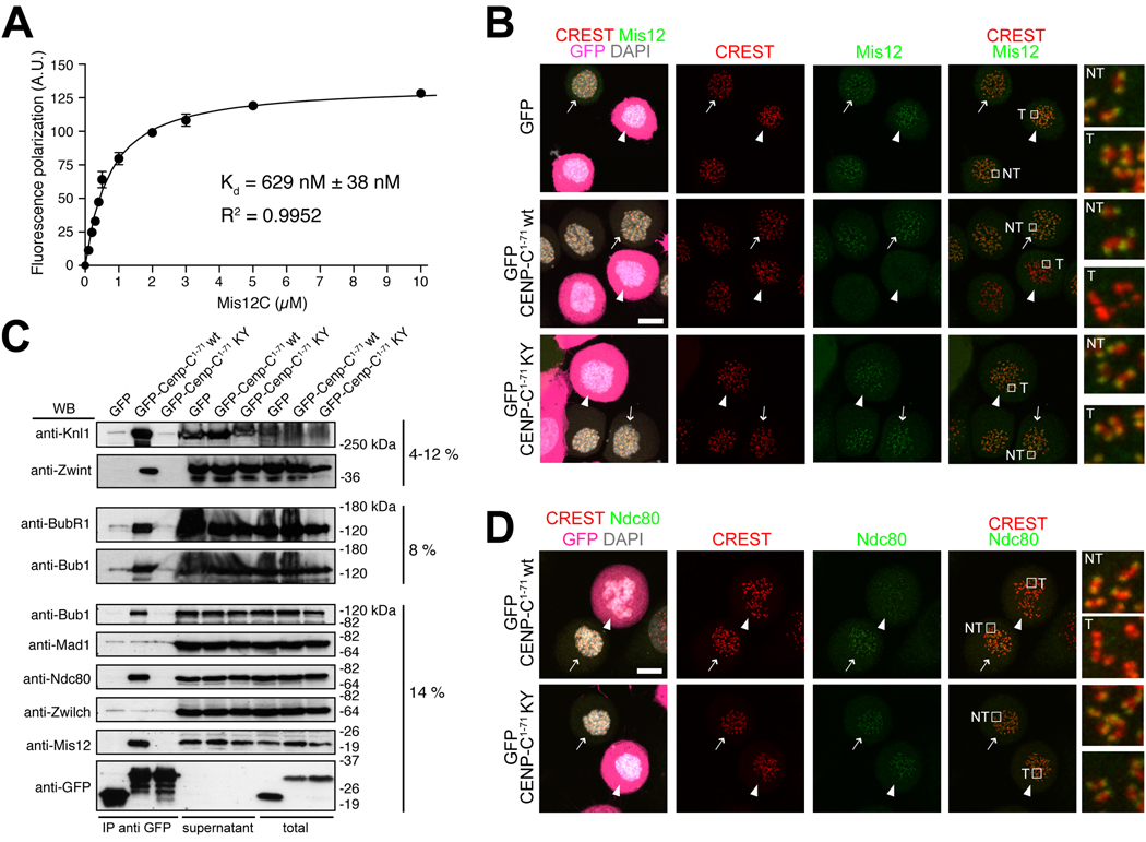
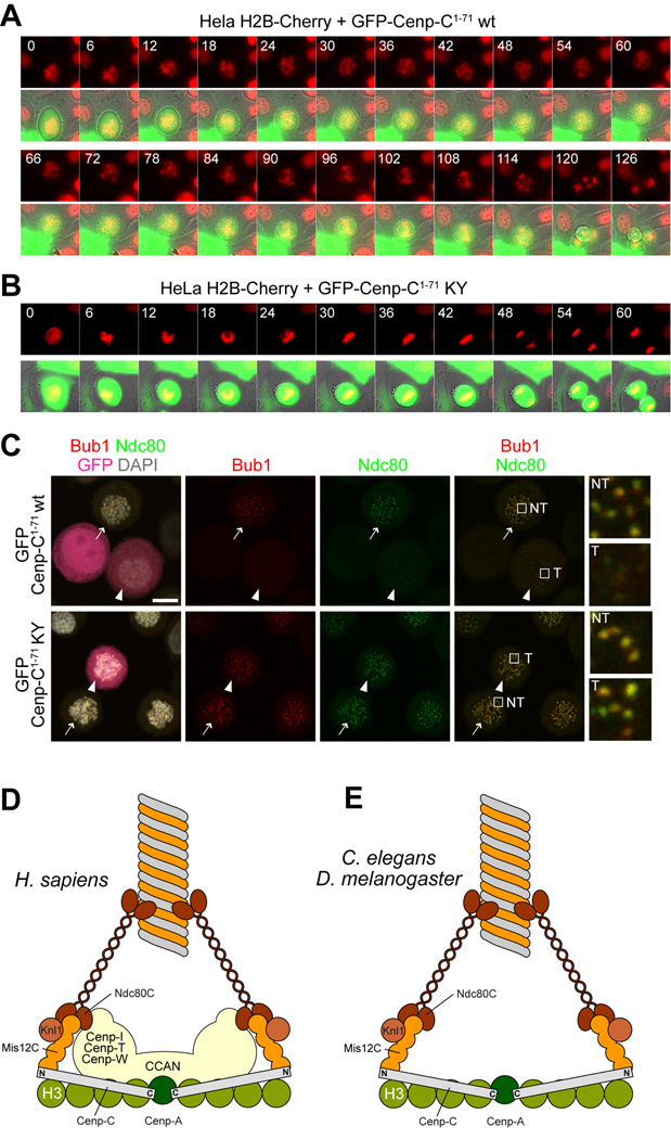
Kinetochores are proteinaceous scaffolds implicated in the formation of load-bearing attachments of chromosomes to microtubules during mitosis. Kinetochores contain distinct chromatin- and microtubule-binding interfaces, generally defined as the inner and outer kinetochore, respectively (reviewed in). The constitutive centromere-associated network (CCAN) and the Knl1-Mis12-Ndc80 complexes (KMN) network are the main multisubunit protein assemblies in the inner and outer kinetochore, respectively. The point of contact between the CCAN and the KMN network is unknown. Cenp-C is a conserved CCAN component whose central and C-terminal regions have been implicated in chromatin binding and dimerization. Here, we show that a conserved motif in the N-terminal region of Cenp-C binds directly and with high affinity to the Mis12 complex. Expression in HeLa cells of the isolated N-terminal motif of Cenp-C prevents outer kinetochore assembly, causing chromosome missegregation. The KMN network is also responsible for kinetochore recruitment of the components of the spindle assembly checkpoint, and we observe checkpoint impairment in cells expressing the Cenp-C N-terminal segment. Our studies unveil a crucial and likely universal link between the inner and outer kinetochore.


Figures

Figure 1
Figure 1. Cenp-C1–400 binds Mis12C
(A) Schematic depiction of the domain organization of human Cenp-C. (B) Size-exclusion chromatography elution profiles and SDS-PAGE analysis of recombinant Mis12CNsl1(1–227) (upper panel), recombinant Cenp-C1–400 (middle panel), and their stoichiometric combination (lower panel). Complex formation is indicated by a shift in the elution profile of Cenp-C1–400 and Mis12CNsl1(1–227) and their appearance, in stoichiometric amounts, in early elution volumes. (C) As in B, but with Mis12CFL-Knl12106–2316 instead of Mis12CNsl1(1–227). The middle panel is the same as in B. (D) Incorporation of the Ndc80Bonsai complex in the Cenp-C1–400-Mis12CNsl1(1–227) complex. The upper panel and lower middle panel are the same as in B.
Figure 2
Figure 2. Electron microscopy analysis of the Cenp-C-Mis12C complex
(A) Representative single particles of human Ndc80C (full length). Scale bar, 50 nm. (B) Representative single particles of Ndc80C-Mis12CNsl1(1–258). (C) Representative single particles of Ndc80C-Mis12CNsl1(1–258)-Cenp-C1–400 (D) Representative field of single particles of the Mis12CNsl1(1-258)-Cenp-C1–400 complex. Particles are indicated by asterisks. Scale bar, 10 nm. (E) Representative field of particles of Mis12CNsl1(1–258). Additional analyses on this complex were recently published [25, 39, 40]. (F) Selected reference-free class averages of the Mis12CNsl1(1–258)-Cenp-C1–400 complex. The complex appears as a linear rod, generally with a prominent bump at one end. Scale bar, 10 nm. (G) Selected reference-free class averages of Mis12CNsl1(1–258) alone. The complex adopts varying degrees of bending, and the globular lobe is often absent or diminished in size. (H) Bending angle analysis of the Mis12 complex in the presence and absence of Cenp-C1–400. The complex is substantially rigidified upon Cenp-C binding.
Figure 3
Figure 3. Wild type but not mutant Cenp-C1–71 binds Mis12C
(A) Size-exclusion chromatography elution profiles and SDS-PAGE analysis of recombinant Mis12CMini (upper panel), recombinant GST-Cenp-C1–71 (middle panel), and their stoichiometric combination (lower panel). Complex formation is indicated by a shift in the elution profile of GST-Cenp-C1–71 and Mis12CMini and their appearance, in stoichiometric amounts, in early elution volumes. (B) Multiple sequence alignment of the N-terminal region of Cenp-CV from the indicated species. The colouring scheme reports the degree of conservation. The position of three predicted helices is shown on the sequence of human Cenp-C. (C) As in A, but with a GST-Cenp-C1–71Y13A mutant. The upper panel is the same as in A. No shift in elution volume is observed, indicating that binding is impaired. (D) As in A, but with a GST-Cenp-C1–71K10A mutant. The upper panel is the same as in A. Also with this mutant no shift in elution volume is observed.
Figure 4
Figure 4. Cenp-C1–71 disrupts outer kinetochore assembly
(A) Fluorescence anisotropy measurements of the binding affinity of Mis12Mini for a synthetic fluorescein-conjugated peptide encompassing residues 1-21 of Cenp-C. (B) Direct fluorescence (GFP, DAPI) or indirect immuno-fluorescence from the indicated species in HeLa cells. In this and subsequent panels, arrows indicate non-transfected GFP-negative cells whereas arrowheads indicate transfected GFP-positive cells. Localization of Mis12 in GFP transfected cells is normal. Mis12 is displaced from kinetochores in cells expressing the wild type sequence of GFP-Cenp-C1–71. The construct localizes in the cytosol but it is not enriched at kinetochores. Mis12 is normally localized in cells expressing the double point mutant GFP-Cenp-C1–71K10A-Y13A mutant (abbreviated as KY). Scale bar = 10 µM. (C) Lysates from HeLa cells expressing the indicated GFP species were used for immuno-precipitations using an anti-GFP antibody. Several outer kinetochore proteins interact with wild type GFP-Cenp-C1–71 but not with its mutated form. Content of IPs from 1.5 mg of cell lysates, 45 µg of supernatants and 45 µg of total cell lysates were loaded on gel. (D) As in B, but detecting Ndc80.
Figure 5
Figure 5. Cenp-C1–71 disrupts the spindle checkpoint
(A) HeLa cells co-expressing histone H2B-Cherry and wild type GFP-Cenp-C1–71 were filmed as they transited through mitosis. Cells attempted congression but failed to align and exited aberrantly, indicative of spindle checkpoint failure. Scale bar = 10 µM. (B) HeLa cells co-expressing histone H2B-Cherry and mutant GFP-Cenp-C1–71-K10A-Y13A divided normally. (C) Direct fluorescence (GFP, DAPI) or indirect immuno-fluorescence from the indicated species in HeLa cells. In this and subsequent panels, arrows indicate non-transfected GFP-negative cells whereas arrowheads indicate transfected GFP-positive cells. Localization of Bub1 and Ndc80 is severely impaired in cells expressing GFP-Cenp-C1–71, whereas it is normal in cells expressing the double point mutant GFP-Cenp-C1–71K10A-Y13A mutant (abbreviated as KY). Scale bar = 10 µM. (D) A model for the interaction of inner and outer kinetochore in mammalian cells. A “two-hand” model predicts that Ndc80 is recruited in part via Mis12C, but also through a pathway that depends on the CCAN. Only the globular C-terminal domain of Knl1 is shown. (E) The CCAN is not present in D. melanogaster or C. elegans, suggesting that the interaction of the Mis12C with Cenp-C is the only contact between the inner and outer kinetochore.>

Comment in

References

    1. Santaguida S, Musacchio A. The life and miracles of kinetochores. EMBO J. 2009;28:2511–2531. - PMC - PubMed
    1. Trazzi S, Bernardoni R, Diolaiti D, Politi V, Earnshaw WC, Perini G, Della Valle G. In vivo functional dissection of human inner kinetochore protein CENP-C. J Struct Biol. 2002;140:39–48. - PubMed
    1. Trazzi S, Perini G, Bernardoni R, Zoli M, Reese JC, Musacchio A, Della Valle G. The C-terminal domain of CENP-C displays multiple and critical functions for mammalian centromere formation. PLoS One. 2009;4:e5832. - PMC - PubMed
    1. Cohen RL, Espelin CW, De Wulf P, Sorger PK, Harrison SC, Simons KT. Structural and functional dissection of Mif2p, a conserved DNA-binding kinetochore protein. Mol Biol Cell. 2008;19:4480–4491. - PMC - PubMed
    1. Carroll CW, Milks KJ, Straight AF. Dual recognition of CENP-A nucleosomes is required for centromere assembly. J Cell Biol. 2010;189:1143–1155. - PMC - PubMed

Publication types

MeSH terms

Substances