

Magnetoreception in an avian brain in part mediated by inner ear lagena
- PMID: 21353559
- PMCID: PMC3062271
- DOI: 10.1016/j.cub.2011.01.058
Magnetoreception in an avian brain in part mediated by inner ear lagena
Abstract
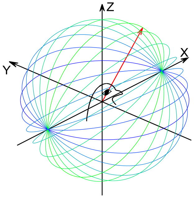
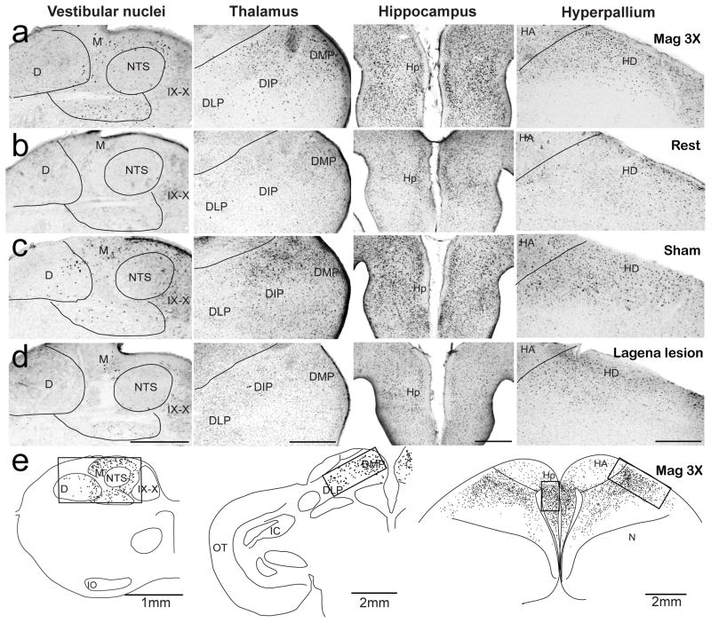
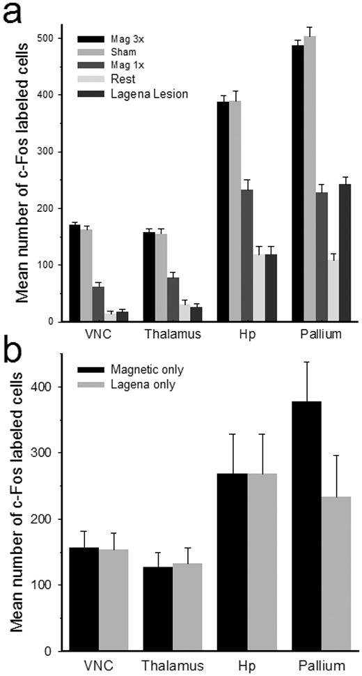
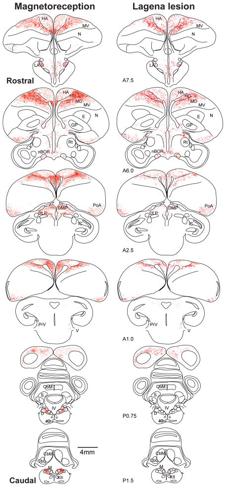
Many animals use the Earth's geomagnetic field for orientation and navigation, but the neural mechanisms underlying that ability remain enigmatic. Support for at least two avian magnetoreceptors exists, including magnetically activated photochemicals in the retina and ferrimagnetic particles in the beak. The possibility of a third magnetoreceptor in the inner ear lagena organs has been suggested. The brain must process magnetic receptor information to derive constructs representing directional heading and geosurface location. Here, we used the c-Fos transcription factor, a marker for activated neurons, to discover where in the brain computations related to a specific set of magnetic field stimulations occur. We found that neural activations in discrete brain loci known to be involved in orientation, spatial memory, and navigation may constitute a major magnetoreception pathway in birds. We also found, through ablation studies, that much of the observed pathway appears to receive magnetic information from the pigeon lagena receptor organs.
Copyright © 2011 Elsevier Ltd. All rights reserved.
Figures
Similar articles
-
Neural correlates of a magnetic sense.Science. 2012 May 25;336(6084):1054-7. doi: 10.1126/science.1216567. Epub 2012 Apr 26. Science. 2012. PMID: 22539554
-
A magnetic pulse does not affect homing pigeon navigation: a GPS tracking experiment.J Exp Biol. 2013 Jun 15;216(Pt 12):2192-200. doi: 10.1242/jeb.083543. Epub 2013 Mar 7. J Exp Biol. 2013. PMID: 23470658
-
No evidence for a magnetite-based magnetoreceptor in the lagena of pigeons.Curr Biol. 2019 Jan 7;29(1):R14-R15. doi: 10.1016/j.cub.2018.11.032. Curr Biol. 2019. PMID: 30620907
-
[Magnetoreception systems in birds: a review of current research].Zh Obshch Biol. 2014 Mar-Apr;75(2):104-23. Zh Obshch Biol. 2014. PMID: 25490840 Review. Russian.
-
Magnetoreception and baroreception in birds.Dev Growth Differ. 2013 Jan;55(1):188-97. doi: 10.1111/dgd.12025. Epub 2012 Dec 17. Dev Growth Differ. 2013. PMID: 23253017 Review.
Cited by
-
Myths in magnetosensation.iScience. 2022 May 23;25(6):104454. doi: 10.1016/j.isci.2022.104454. eCollection 2022 Jun 17. iScience. 2022. PMID: 35677648 Free PMC article. Review.
-
Odours stimulate neuronal activity in the dorsolateral area of the hippocampal formation during path integration.Proc Biol Sci. 2014 Mar 26;281(1783):20140025. doi: 10.1098/rspb.2014.0025. Print 2014 May 22. Proc Biol Sci. 2014. PMID: 24671977 Free PMC article.
-
Zebrafish and medaka offer insights into the neurobehavioral correlates of vertebrate magnetoreception.Nat Commun. 2018 Feb 23;9(1):802. doi: 10.1038/s41467-018-03090-6. Nat Commun. 2018. PMID: 29476093 Free PMC article.
-
Semicircular canal biomechanics in health and disease.J Neurophysiol. 2019 Mar 1;121(3):732-755. doi: 10.1152/jn.00708.2018. Epub 2018 Dec 19. J Neurophysiol. 2019. PMID: 30565972 Free PMC article. Review.
-
Extracellular recordings reveal absence of magneto sensitive units in the avian optic tectum.J Comp Physiol A Neuroethol Sens Neural Behav Physiol. 2014 Dec;200(12):983-96. doi: 10.1007/s00359-014-0947-6. Epub 2014 Oct 4. J Comp Physiol A Neuroethol Sens Neural Behav Physiol. 2014. PMID: 25281335 Free PMC article.
References
-
- Lohmann KL. Magnetic-field perception. Nature. 2010;464:1140–1142. - PubMed
-
- Wiltschko R, Wiltschko W. Magnetic orientation and magnetoreception in birds and other animals. J Comp Physiol. 2005;191:675–693. - PubMed
-
- Möller A, Sagasser S, Wiltschko W, Schierwater B. Retinal cryptochrome in a migratory passerine bird: a possible transducer for the avian magnetic compass. Naturwissenschaften. 2004;91:585–588. - PubMed
-
- Fleissner G, Stahl B, Thalau P, Falkenberg G, Fleissner G. A novel concept of Fe-mineral-based magnetoreception: histological and physicochemical data from the upper beak of homing pigeons. Naturwissenschaften. 2007;94:631–642. - PubMed
Publication types
MeSH terms
Substances
Grants and funding
LinkOut - more resources
Full Text Sources
