Review
. 2011 Apr;17(4):197-206.
doi: 10.1016/j.molmed.2010.12.005. Epub 2011 Feb 23.

Optogenetic investigation of neural circuits in vivo

Affiliations
Review

Optogenetic investigation of neural circuits in vivo

Matthew E Carter et al. Trends Mol Med. 2011 Apr.

Abstract

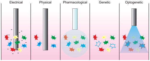
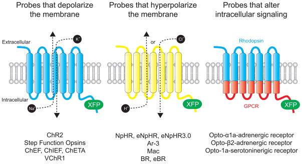
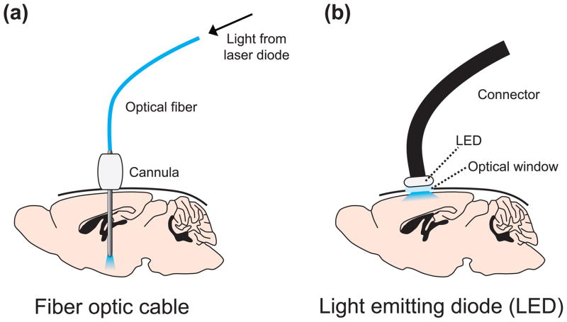
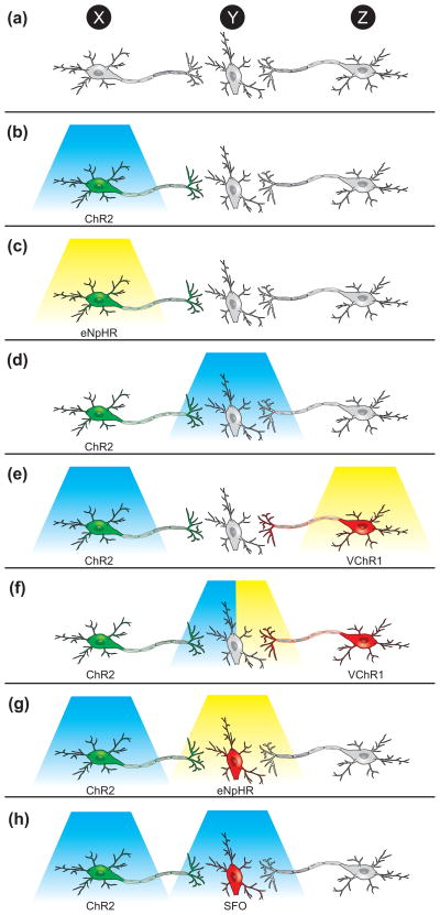
The recent development of light-activated optogenetic probes allows for the identification and manipulation of specific neural populations and their connections in awake animals with unprecedented spatial and temporal precision. This review describes the use of optogenetic tools to investigate neurons and neural circuits in vivo. We describe the current panel of optogenetic probes, methods of targeting these probes to specific cell types in the nervous system, and strategies of photostimulating cells in awake, behaving animals. Finally, we survey the application of optogenetic tools to studying functional neuroanatomy, behavior and the etiology and treatment of various neurological disorders.


Figures

Figure 1
Figure 1
Strategies to manipulate neural activity in vivo. Cells in the nervous system reside in heterogeneous populations composed of different subtypes (here depicted as different colors). Electrical microstimulation techniques have high temporal precision but affect all cells and fibers surrounding the electrode. Physical probes irreversibly ablate or reversibly cool cells to reduce neural activity, but also affect all cells and fibers surrounding the electrode. Pharmacological injection of a drug might be able to target a particular cell-type based on cell-specific protein expression, but the drug could remain in the system for minutes or hours after injection. Genetic inactivation of specific cells (such as the cells depicted in blue) also lacks temporal precision and is often irreversible. Optogenetic stimulation or inhibition of cells using light allows for cell-type specific targeting of optogenetic probes (such as the cells depicted in blue) with millisecond-timescale precision of activation.
Figure 2
Figure 2
Optogenetic probes. All optogenetic probes are based on opsins, seven transmembrane domain proteins that interact with a chromophore (retinal or vitamin A) to become light sensitive. Probes that depolarize the membrane act as nonspecific cation channels that open in response to pulses of light. Probes that hyperpolarize the membrane actively pump either Cl ions (in the case of NpHRs) or protons in response to pulses of light. Probes that alter intracellular activity are chimeric proteins composed of rhodopsin in the extracellular and transmembrane domains and a GPCR in the intracellular domain.
Figure 3
Figure 3
Strategies to deliver light to transduced neurons in vivo. (a) A guide cannula is stereotaxically implanted above a target region for subsequent placement of an optic fiber. The fiber is attached to a laser diode, which might be attached to a computer or pulse generator for automatic stimulation protocols. (b) An optical window is stereotaxically implanted above a target region. A light-emitting diode (LED) is placed above the window to illuminate surface neurons and is connected to a computer for automatic stimulation protocols.
Figure 4
Figure 4
Optogenetic strategies to investigate neural circuits in vivo. (a) A schematic of a neural circuit in which a population of neurons, Population X, projects monosynaptically to Population Y, which also receives projections from Population Z. (b) To determine the effect of stimulating X on behavior, Population X neurons could be transduced with ChR2 (or a variant) and stimulated with blue light. The effect of stimulating X on neural activity in Y could be determined by simultaneously placing an electrode in Y. (c) To determine the effect of inhibiting X on behavior, Population X could be transduced with eNpHR (or another inhibitory probe) and stimulated with yellow light. The effect of inhibiting X on neural activity in Y could be determined by simultaneously placing an electrode in Y. (d) To determine the effect of stimulating the projections of X onto Y on behavior or neural activity in Y, Population X could be transduced with ChR2 and light could be delivered to Y. The blue light will only activate ChR2 on the distal projections and thus the role of the specific synaptic connections can be probed. (e) To determine the relative contributions of activity in X or Z on behavior or activity in Y, Population X could be transduced with ChR2, Population Z could be transduced with VChR1, and X could be stimulated with blue light and Z with yellow light (f) To more specifically determine the relative contributions of activity in X or Z on activity in Y, Population X could be transduced with ChR2, Population Z could be transduced with VChR1, and the projections onto Y could be stimulated with either blue or yellow light while recording with an optrode. (g) To determine the necessity of Y on the ability of X to influence behavior, Population X could be transduced with ChR2, Population Y could be transduced with eNpHR, and X could be stimulated with blue light while Y is inhibited with yellow light. (h) To determine the effect of turning up the gain of activity in Y while stimulating X on behavior, Population X could be transduced with ChR2, and Population Y could be transduced with a step-function opsin (SFO) that converts a single light pulse into a graded increase in membrane potential. Then X could be stimulated while Y is stimulated to subthreshold potential with a single pulse of blue light.

References

    1. Luo L, et al. Genetic dissection of neural circuits. Neuron. 2008;57:634–660. - PMC - PubMed
    1. Carter M, Shieh JC. Guide to research techniques in neuroscience. Elsevier/Academic Press; 2010.
    1. Deisseroth K, et al. Next-generation optical technologies for illuminating genetically targeted brain circuits. J Neurosci. 2006;26:10380–10386. - PMC - PubMed
    1. Boyden ES, et al. Millisecond-timescale, genetically targeted optical control of neural activity. Nat Neurosci. 2005;8:1263–1268. - PubMed
    1. Zhang F, et al. Red-shifted optogenetic excitation: a tool for fast neural control derived from Volvox carteri. Nat Neurosci. 2008;11:631–633. - PMC - PubMed

Publication types