Review
. 2011 Apr;34(4):198-209.
doi: 10.1016/j.tins.2011.01.005. Epub 2011 Feb 25.

Cytoprotective protein C pathways and implications for stroke and neurological disorders

Affiliations
Review

Cytoprotective protein C pathways and implications for stroke and neurological disorders

Berislav V Zlokovic et al. Trends Neurosci. 2011 Apr.

Abstract

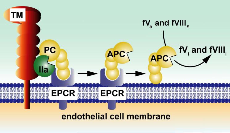
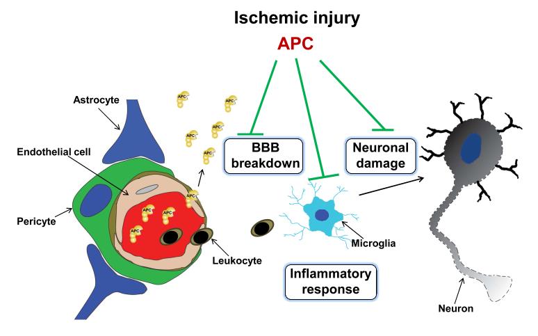
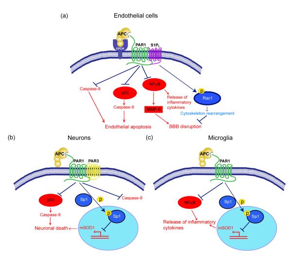
Recent studies indicate that single-action-single-target agents are unlikely to cure CNS disorders sharing a pathogenic triad consisting of vascular damage, neuronal injury/neurodegeneration and neuroinflammation. Here we focus on a recent example of a multiple-action-multiple-target approach for CNS disorders based on newly discovered biological properties of activated protein C (APC), an endogenous plasma protease with antithrombotic, cytoprotective and anti-inflammatory activities in the CNS. We propose that APC-mediated signaling through the protease activated receptor-1 (PAR1) can favorably regulate multiple pathways within the neurovascular unit in non-neuronal cells and neurons during acute or chronic CNS insults, leading to stabilization of the blood-brain barrier (BBB), neuroprotection and control of neuroinflammation. Although much remains to be understood regarding the biology of APC, preclinical studies suggest that APC has promising applications as disease-modifying therapy for ischemic stroke and other neuropathologies whose underlying pathology involves deficits in the vasculo-neuronal-inflammatory triad.


Figures

Figure 1
Figure 1. Pathways for protein C activation and expression of anticoagulant activity
The endothelial cell receptors, thrombomodulin (TM) and EPCR, are required for efficient activation of protein C by alpha-thrombin (IIa). Dissociation of APC from EPCR allows for the expression of APC’s anticoagulant activity, whereas retention of APC bound to EPCR allows APC to express multiple direct cellular activities in the endothelium (see Figure 3a). APC conveys its anticoagulant activity when bound to cell membrane surfaces by cleaving the activated cofactors Va (fVa) and VIIIa (fVIIIa) to yield the inactivated cofactors, fVi and fVIIIi. Various proteins such as plasma protein S and factor V, high density lipoprotein particles, lipids (e.g., negatively charged phosphatidyl serine and cardiolipin, phosphatidyl ethanolamine) and certain glycosphingolipids (e.g., glycosyl ceramide, lactosyl ceramide, etc) provide APC-cofactor activities that accelerate proteolysis of factors Va and VIIIa by APC (not shown). Figure adapted from a review article originally published by L. Mosnier, B. Zlokovic and J.H. Griffin (2007) The cytoprotective protein C pathway Blood, 109, 3161-3172 [28].
Figure 2
Figure 2. Control of ischemic injury to the neurovascular unit by APC and/or its cell-signaling analogs
APC protects the BBB integrity and ameliorates post-ischemic BBB breakdown thus preventing secondary neuronal damage mediated by entry of several blood-derived neurotoxic and vasculotoxic molecules. It crosses an intact BBB via EPCR-dependent transport to reach its neuronal targets in brain and expresses its direct neuronal protective activity to prevent neuronal damage. APC also expresses anti-inflammatory activities by blocking early post-ischemic infiltration of brain by neutrophils. It also suppresses microglia activation. The key membrane signaling receptor mediating beneficial effects of APC on different cell types within the neurovascular unit is PAR1, as illustrated in Figure 3.
Figure 3
Figure 3. APC protective signaling activities in different cell types within the neurovascular unit
a. BBB ‘sealing’ effect and direct vasculoprotection: Beneficial cytoprotective activities of APC and its cytoprotective-selective variants involve direct effects on endothelial cells that require the cellular receptors EPCR and PAR-1 and EPCR-dependent PAR1-mediated cross-activation of sphingosine 1-phosphate receptor 1 (S1P1). Cross-activation of S1P1 activates Rac1 leading to Rac1-dependent stabilization of the cytoskeleton, which results in enhanced integrity of the endothelial membranes (right). APC also suppresses NF-kB-dependent transcriptional activation of MMP-9 which in turn blocks degradation of the BBB basement membrane, thereby preventing intracerebral bleeding (middle right). By controlling Nf-κB nuclear translocation, APC blocks expression of pro-inflammatory cytokines, thereby limiting neuroinflammation (middle left). By activating PAR-1 in a EPCR-dependent manner, APC suppresses the pro-apoptotic p53 transcription factor and p53-dependent transcription of Bax (not shown) and directly upregulates anti-apoptotic Bcl-2 (not shown), which results in blockade of caspase-9 activation and controls the intrinsic apoptotic pathway (middle left). APC can also block the extrinsic apoptotic pathway and caspase-8 activation (left). b. Direct neuronal protection: APC and its cytoprotective cell signaling variants act through PAR-1 and PAR-3 to inhibit p53 activation in injured neurons resulting in blockade of caspase-9-dependent intrinsic apoptotic pathway (left) and caspase-8-dependent extrinsic apoptotic pathway (right). APC also blocks nuclear transport of the transcription factor, Sp1, through phosphorylation of cytoplasmic Sp1 resulting in transcriptional suppression of mutant SOD1 (mSOD1) expression in motor neurons in a mouse ALS model (middle). c. Anti-inflammatory activity: In SOD1 mutant mice, APC and its cell signaling variants downregulate mSOD1 expression though PAR-1 and the downstream mechanisms as noted in panel b. This suppresses microglia activation resulting in reduced number of activated microglia as well as blockade of inflammatory cytokine production from microglia (right). On the left is shown that APC blocks NF-κB activation in microglia providing another anti-inflammatory pathway mediated by a blockade of NF-kB-dependent transcriptional expression of different pro-inflammatory cytokines as noted above in a. for endothelial cells.
Figure I
Figure I. APC exosite structures responsible for APC activities
a. The model of full-length APC is based on the serine protease domain crystal structure of APC (,; Protein Data Bank entry 1AUT; http://www.rcsb.org/pdb/explore/explore.do?structureId=1AUT). In the ribbon diagram of the polypeptide structure of APC, the folded serine protease domain at the top of panel, the two EGF-like domains and the N-terminal Gla-domain (which mediates binding to phospholipid membranes and EPCR) are shown in gray. The active serine protease site is in red. The positively charged lysine residues within the so-called 37 loop (KKK191-193) and the arginine residues in the calcium binding-binding loop (RR229/230) determine APC specificity for factors Va and VIIIa. b. Amino acid residues that determine exosite specificity of the recombinant wild-type APC (wt-APC) protease domain are schematically identified in the space filling model where the close apposition of the 37-loop and the calcium-binding loop positively charged residues (blue) comprising lysine residues K191-193, and arginine residues R229 and R230 are seen inside the yellow rectangle. The active site triad of serine, histidine and aspartic acid residues characteristic of serine proteases is shown in green while red indicates negative side chains. These five basic residues are critical for recognition of the coagulation factor Va and thus for anticoagulant activity; however, they are not required for normal anti-apoptotic activity. The 5A-APC mutant (containing 5 Ala substitutions at residues 191-193 and 229-230) is depicted on the right side inside the yellow rectangle showing a remarkable loss of the positively charged amino acid side chain cluster. The model of wt-APC is based on the serine protease domain structure of APC 1AUT (107) and was generated using Modeller (107).

Similar articles

Cited by

References

    1. Moskowitz MA, et al. The science of stroke: mechanisms in search of treatments. Neuron. 2010;67:181–198. - PMC - PubMed
    1. Guo S, Lo EH. Dysfunctional cell-cell signaling in the neurovascular unit as a paradigm for central nervous system disease. Stroke. 2009;40:S4–S7. - PMC - PubMed
    1. Zlokovic BV. The blood-brain barrier in health and chronic neurodegenerative disorders. Neuron. 2008;57:178–201. - PubMed
    1. Iadecola C. The overlap between neurodegenerative and vascular factors in the pathogenesis of dementia. Acta Neuropathol. 2010;120:287–296. - PMC - PubMed
    1. Zlokovic BV. Neurodegeneration: are we overlooking the role of neurovascular unit? Nat. Med. 2010;16:1370–1371. - PubMed

Publication types

MeSH terms