Review
. 2011 May;19(5):225-32.
doi: 10.1016/j.tim.2011.01.007. Epub 2011 Feb 25.

A play in four acts: Staphylococcus aureus abscess formation

Affiliations
Review

A play in four acts: Staphylococcus aureus abscess formation

Alice G Cheng et al. Trends Microbiol. 2011 May.

Abstract

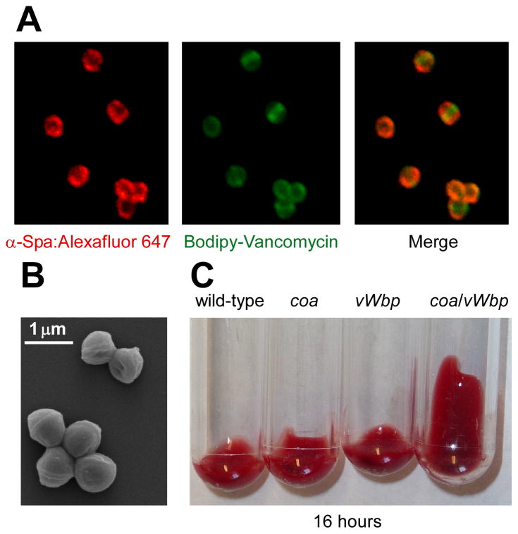
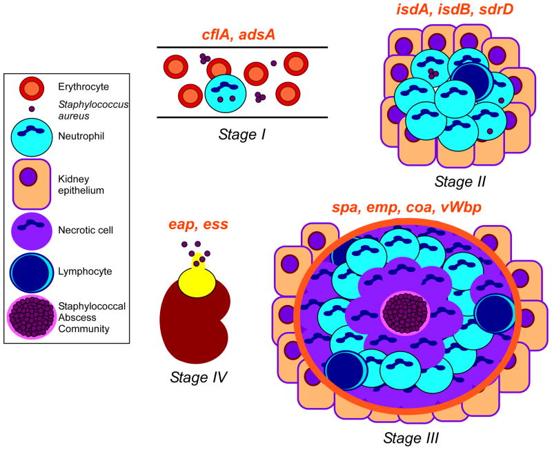
Staphylococcus aureus is an important human pathogen that causes skin and soft tissue abscesses. Abscess formation is not unique to staphylococcal infection and purulent discharge has been widely considered a physiological feature of healing and tissue repair. Here we present a different view, whereby S. aureus deploys specific virulence factors to promote abscess lesions that are distinctive for this pathogen. In support of this model, only live S. aureus is able to form abscesses, requiring genes that act at one or more of four discrete stages during the development of these infectious lesions. Protein A and coagulases are distinctive virulence attributes for S. aureus, and humoral immune responses specific for these polypeptides provide protection against abscess formation in animal models of staphylococcal disease.


Conflict of interest statement

Conflict of Interest

Alice Cheng, Andrea DeDent, Olaf Schneewind and Dominique Missiakas are named inventors on a patent owned by The University of Chicago, which is the subject of a license agreement with Novartis Vaccines and Diagnostics.

Figures

Figure 1
Figure 1
Staphylococcus aureus characteristics and distinguishing features. (a) The immunoglobulin binding properties of staphylococcal protein A were revealed by confocal laser scanning immunofluoresence microscopy as previously described . Alexafluor-647 conjugated immunoglobulin (red) binds to protein A (left), Bodipy-conjugated vancomycin (green) stains staphylococcal peptidoglycan (middle), and a merger of the two data sets reveals the deposition of immunoglobulin on the surface of staphylococci (right). (b) Shape and clustering of S. aureus cells imaged by scanning electron microscopy (SEM; image courtesy of Matt Frankel, University of Chicago). (c) S. aureus coagulates blood, a feature conferred by two secreted coagulases: staphylocoagulase (Coa) and von Willebrand factor binding protein (vWbp). Lepirudin anti-coagulated mouse blood was incubated with 105 cells of wild-type S. aureus Newman or isogenic mutants lacking coa, vWbp, or coa/vWbp and examined over time for the formation of a blood clot. Blood containing staphylococci lacking both coagulase genes do not form a clot following a 16 hour observation period.
Figure 2
Figure 2
Working model for staphylococcal abscess formation and persistence in host tissues. Stage I: following intravenous inoculation, S. aureus survives in the bloodstream and disseminates via the vasculature to peripheral organ tissues. Stage II: staphylococci in renal tissue attract a massive infiltrate of polymorphonuclear leukocytes and other immune cells. Stage III: abscesses mature showing a central accumulation of the pathogen (SAC) surrounded by a pseudocapsule of fibrin deposits (pink rim), and zones of necrotic and healthy polymorphonuclear neutrophils (PMNs; purple and light blue cells, respectively), and finally a rim of eosinophilic material (orange rim). Stage IV: abscesses mature and rupture on the organ surface to initiate new rounds of infections. Genes required for specific stages of staphylococcal abscess development are in red above the corresponding stage of infection. Figure adapted with the authors’ permission from an article published by Cheng and colleagues.
Figure 3
Figure 3
Monitoring and visualizing Staphylococcus aureus abscess formation. (a)–(e) Visualization of lesions in kidneys following infection at Stages II, III and IV as previously described [14]. Paraffin embedded kidneys were thin-sectioned, stained with hematoxylineosin, mounted on glass slides and examined by light microscopy for the formation of staphylococcal abscesses. Micrographs on the left (a, c, e) show sections of entire kidneys. Micrographs on the right (b, d) represent magnifications of the area framed in the white box in (a) and (c). (b) Stage II: Site of inflammation with immune cell infiltrates (green arrowhead). (d) Stage III: staphylococci are clearly distinguishable as central nidus, staphylococcal abscess community (SAC), within the maturing abscess (yellow arrowheads). The SAC is surrounded by an amorphous, eosinophilic capsule (see Figure 4) followed by a zone of dead PMNs (polymorphonuclear neutrophils; white box), apparently healthy PMNs (red box), and necrotic PMNs (green box). The entire abscess region is separated from healthy kidney tissue by a second eosinophilic layer, which appears to increase in size over time. (e) Stage IV: Abscesses migrate to the periphery of organ tissue. The organization of SACs and immune cells during Stage III is abolished prior to rupture of the lesion onto the organ surface. (f) Representative distribution of bacteria in blood (dashed line) and kidney tissue (solid line) over time following intravenous infection of mice with 1 × 10⁷ colony forming units (CFU) of S. aureus. Bacterial loads are represented as CFU per g of blood or renal tissue over time following infection.
Figure 4
Figure 4
SACs during Stage III. At the center of an abscess lesion, S. aureus cells are found as tightly associated mass covered with a granular, electron-dense substance. Bacteria are enclosed by an amorphous pseudocapsule (white arrowheads) that separates the SAC from neighboring immune cells. Image was captured from the kidney of an animal infected with S. aureus Newman. Organ tissue was thin sectioned, fixed, dehydrated and sputter-coated with platinum/palladium and analyzed by scanning electron microscopy.

References

    1. Kloos W, et al. The genus Staphylococcus. In: Balows A, et al., editors. The prokaryotes. Springer-Verlag; 1992. pp. 1369–1420.
    1. Götz F, et al. The Genera Staphylococcus and Macrococcus. In: Dworkin M, et al., editors. The Prokaryotes. Springer; New York: 2006. pp. 5–75.
    1. Lowy FD. Staphylococcus aureus infections. New Engl J Med. 1998;339:520–532. - PubMed
    1. Brumfitt W, Hamilton-Miller J. Methicillin-resistant Staphylococcus aureus. N Engl J Med. 1989;320:1188–1199. - PubMed
    1. DeLeo FR, Chambers HF. Waves of resistance: Staphylococcus aureus in the antibiotic era. Nat Rev Microbiol. 2009;7:629–641. - PMC - PubMed

Publication types

MeSH terms