. 2011 Jul;55(1):96-102.
doi: 10.1016/j.jhep.2010.10.020. Epub 2010 Dec 7.

Alpha-fetoprotein-thymidine kinase-luciferase knockin mice: a novel model for dual modality longitudinal imaging of tumorigenesis in liver

Affiliations

Alpha-fetoprotein-thymidine kinase-luciferase knockin mice: a novel model for dual modality longitudinal imaging of tumorigenesis in liver

Xincheng Lu et al. J Hepatol. 2011 Jul.

Abstract

Background & aims: Hepatocellular carcinoma (HCC) is frequently a lethal disease and one of the few malignancies that is still increasing in incidence around the world. Better animal models are highly desired to investigate the molecular basis of HCC and to develop novel therapeutic strategies. Alpha-fetoprotein (Afp) gene is expressed in fetal liver, silenced soon after birth, and highly re-expressed in hepatocellular carcinomas (HCC). We aimed to take advantage of the dramatic re-expression of the Afp gene in HCC to develop a hepatocarcinogenesis reporter (HCR) mouse model for dual-modality, longitudinal in vivo imaging of liver tumor development, and progression.

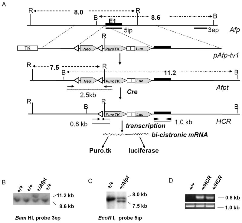
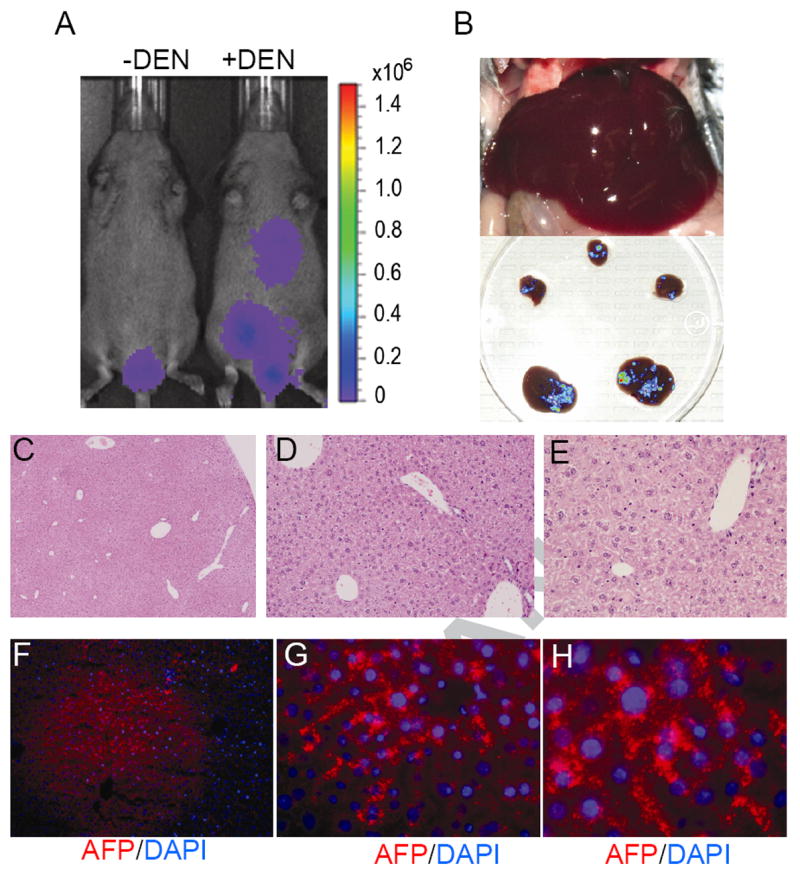
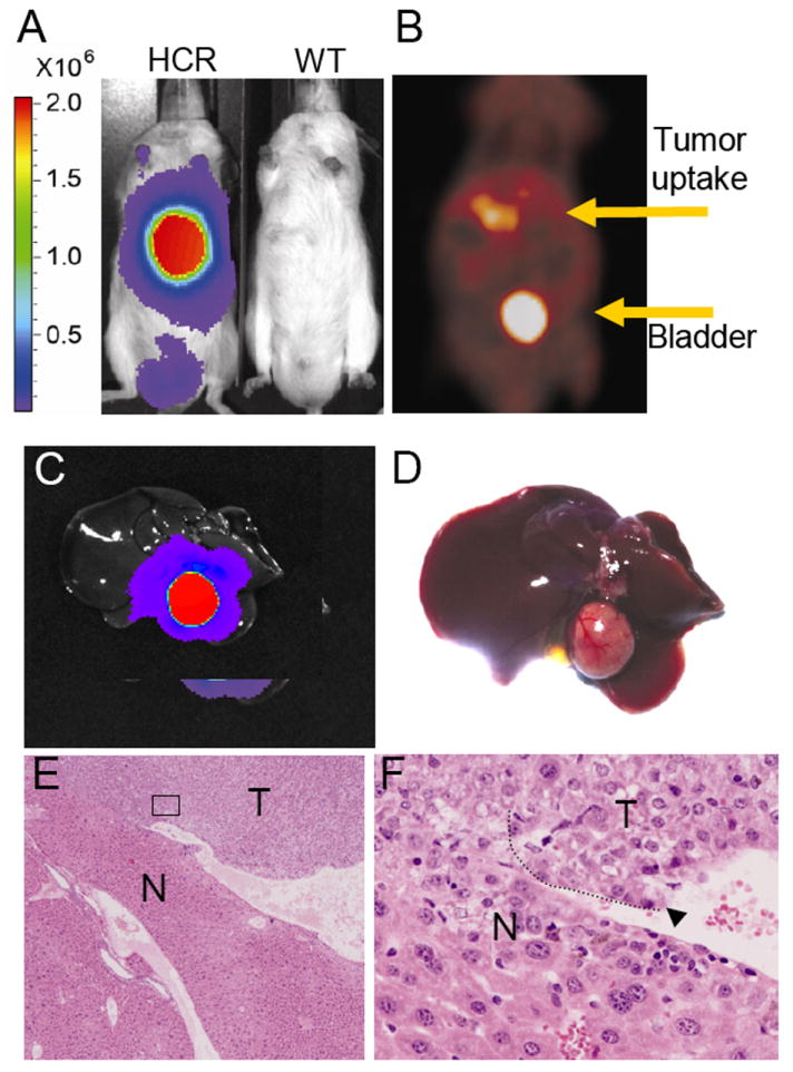
Methods: Knock in mice were established by placing a thymidinekinase (tk)-luciferase (luc) reporter gene cassette under the transcriptional control of the endogenous Afp promoter. DEN, a liver carcinogen, was used to induce liver tumors, which was monitored by both luc-based bioluminescent (BL) and tk-based positron emission tomography (PET) imaging.

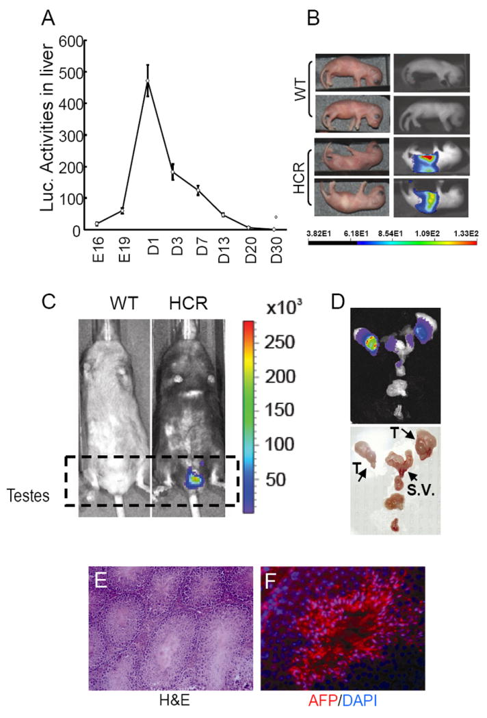
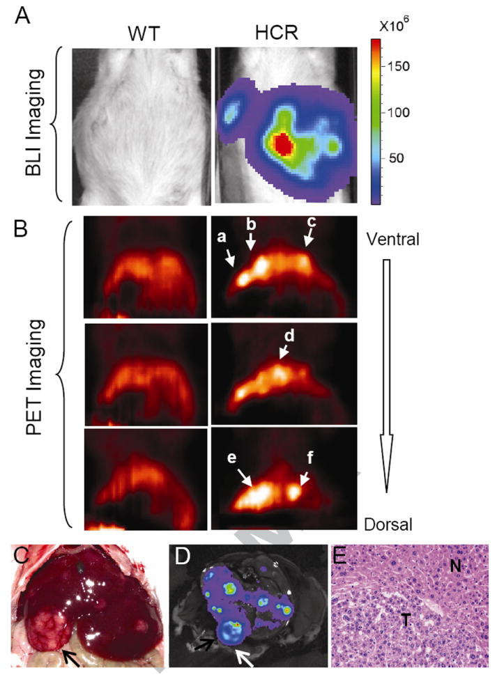
Results: The expression profile of luc was identical to that of the endogenous Afp gene during development. As early as 2 months after the exposure to DEN, BLI revealed multifocal signals in the liver, long before the appearance of histologically apparent neoplastic lesions. By 6 months, BL and PET dual imaging showed strong signals in malignant HCC. By serendipity, a strong BL signal was also detected in adult testes, a previously unknown site of Afp expression.

Conclusions: The HCR model enables longitudinal monitoring of liver tumor development and progression, providing a powerful tool in developing chemoprevention and therapeutic strategies for HCC.


Figures

Figure 1
Figure 1. Gene targeting at the Afp locus
A) A schematic illustration of the gene targeting strategy. Targeting vector (pAfp-tv1) contains a LoxP-Neo-LoxP-PuroTK-ires-Luc cassette between the two arms of homology for gene targeting. By design, the cassette is inserted behind the A of the ATG translation start codon of the Afp gene, generating the primary knock-in allele (the Afpt allele). Cre-mediated deletion of the Neo selection marker then gave rise to the final knock-in allele (the HCR allele). Transcription from this allele, driven by the Afp promoter, is expected to produce a bi-cistronic mRNA with which Puro.TK and luciferase can be produced upon translation, providing two potential reporters for imaging. Targeted clones were identified by Southern blot by the presence of a novel 11.2 Kb Bam HI fragment in addition to the 8.6 Kb wild-type fragment using a 3’ external probe (3ep) and the presence of a 7.5 Kb Eco RI fragment in addition to the 8.0 wild-type fragment by 5’ internal probe (5ip). The HCR allele was detected by a PCR genotyping strategy using a pair of primer corresponding to the Afp promoter and the Puro.TK cassette, respectively. This PCR is designed to generate a 2.5 kb and 0.8 kb product from the afp-t and HCR allele respective (the 2.5 kb product is not amplified under our experimental condition). The sizes of the diagnostic Bam HI (B) and Eco RI (E) fragments of wild-type (Afp) and targeted (Afpt) allele, respectively, were indicated. The positions of the pair of primers used to identify the HCR allele are also shown. B) Initial screening for putative clones containing the afp-t allele by Southern blot. C) Identification of correctly targeted clones by Southern blot from the putative targeted clones identified in B. D) Identification of mice carrying the HCR allele by PCR genotyping. HCR mice were identified by detecting the 0.8 kb PCR fragment specific for the HCR allele (upper panel). As a control, another pair of primers was used to amplify a 1.0 kb fragment from the wild-type Afp gene (lower panel).
Figure 2
Figure 2. Detection of luciferase activity in HCR mice
A) Detection of luciferase activity in the liver of HCR mice at various ages by chemoluminescent assay. B) Detection of luciferase activity in newborn HCR mice by bioluminescent imaging. C) Detection of luciferase activity in adult HCR mice. Note the pattern of luciferase activity in testes. D) BLI of a male reproductive tract. Abbreviations used are: T, testes; S.V., seminal vesicles. E) Photographic image of reproductive tract. F) Histology of testes (magnification 20 ×). G) Detection of Afp in testes by immunofluorescence (magnification 40 ×).
Figure 3
Figure 3. Visualization of early preneoplastic lesions in HCR mice by BLI
A) BLI on a pair of HCR mice. The animal on the right was injected with a single dose of DEN at two weeks of age and imaged two months later. The animal on the left was injected with vehicle control. Note the detection of hepatic BLI signal only in the DEN-injected animal. The testicular signals varied from animal to animal and were not correlated with DEN treatment (not shown). B) A photograph of the liver showing normal gross morphology. C) Ex vivo BLI of dissected liver lobes from DEN injected mouse superimposed on a digital photograph. D, E) Hemotoxylin and eosin staining of liver sections from DEN injected mouse (magnification, D, 5 ×; E, 20×). F, G). Expression of Afp in DEN-treated mouse liver detected by immunofluorescence (red, Afp. blue, DAPI. Magnification, F, 10 ×; G, 40 ×).
Figure 4
Figure 4. BLI and micro-PET dual modality imaging of liver tumors mice six months after DEN treatment
A) BLI of an HCR mouse (right) together with a wild type control (left). Mice were treated as described in Fig. 3 legend. Note the focal nature of BLI signals. B) Micro-PET imaging of the same mice. Serial coronal sections from ventral to dorsal were collected, and three sections are presented. Note distinct patterns of PET signals detected at different depths from the ventral surface of the liver. C) A photograph showing multiple visible liver tumors upon autopsy (top), most of which were Luc-positive based on ex vivo BLI as shown in D. Arrows point to a large tumor visible by both visual inspection and BLI. E) H&E analysis of a typical tumor. Most tumors were classified as benign hepatomas at this stage.
Figure 5
Figure 5. BLI and micro-Pet imaging of malignant HCC
Mice were treated with DEN and imaged nine months later to allow sufficient time for tumor progression. A) BLI of wild type and HCR mice in vivo as described in Fig. 3A. The HCR mouse was also subject to micro-PET imaging (B). A single tumor was identified upon autopsy that was highly positive by ex vivo BLI (C, D). E, F) H&E staining of tumor sections. Note highly heterogeneous nuclear morphology and extensive invasion of tumor cells into surrounding normal liver tissues.

Comment in

  • Focus.
    Friedman SL. Friedman SL. J Hepatol. 2011 Jul;55(1):1-2. doi: 10.1016/j.jhep.2011.03.001. Epub 2011 Mar 22. J Hepatol. 2011. PMID: 21396971 No abstract available.

References

    1. Thorgeirsson SS, Grisham JW. Molecular pathogenesis of human hepatocellular carcinoma. Nat Genet. 2002 Aug;31(4):339–346. - PubMed
    1. Block TM, Mehta AS, Fimmel CJ, Jordan R. Molecular viral oncology of hepatocellular carcinoma. Oncogene. 2003 Aug 11;22(33):5093–5107. - PubMed
    1. Varela M, Sanchez W, Bruix J, Gores GJ. Hepatocellular carcinoma in the setting of liver transplantation. Liver Transpl. 2006 Jul;12(7):1028–1036. - PubMed
    1. Ahmad I, Sansom OJ, Leung HY. Advances in mouse models of prostate cancer 3. Expert Rev Mol Med. 2008;10:e16. - PubMed
    1. Kim CF, Jackson EL, Kirsch DG, Grimm J, Shaw AT, Lane K, et al. Mouse models of human non-small-cell lung cancer: raising the bar. Cold Spring Harb Symp Quant Biol. 2005;70:241–250. - PubMed

Publication types

MeSH terms