Review
. 2011 May;59(5):784-96.
doi: 10.1016/j.eururo.2011.02.033. Epub 2011 Feb 23.

Shock wave technology and application: an update

Affiliations
Review

Shock wave technology and application: an update

Jens J Rassweiler et al. Eur Urol. 2011 May.

Abstract

Context: The introduction of new lithotripters has increased problems associated with shock wave application. Recent studies concerning mechanisms of stone disintegration, shock wave focusing, coupling, and application have appeared that may address some of these problems.

Objective: To present a consensus with respect to the physics and techniques used by urologists, physicists, and representatives of European lithotripter companies.

Evidence acquisition: We reviewed recent literature (PubMed, Embase, Medline) that focused on the physics of shock waves, theories of stone disintegration, and studies on optimising shock wave application. In addition, we used relevant information from a consensus meeting of the German Society of Shock Wave Lithotripsy.

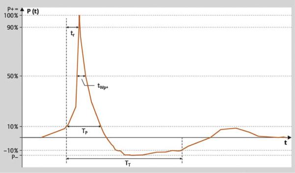
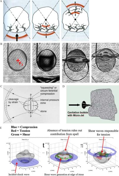
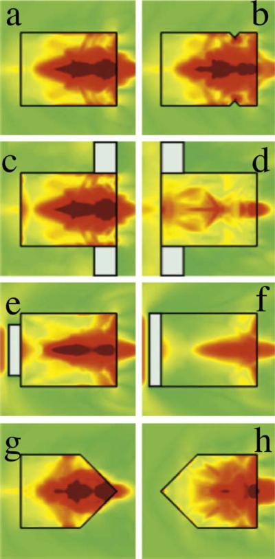
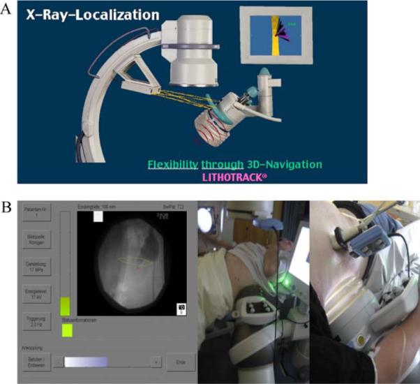
Evidence synthesis: Besides established mechanisms describing initial fragmentation (tear and shear forces, spallation, cavitation, quasi-static squeezing), the model of dynamic squeezing offers new insight in stone comminution. Manufacturers have modified sources to either enlarge the focal zone or offer different focal sizes. The efficacy of extracorporeal shock wave lithotripsy (ESWL) can be increased by lowering the pulse rate to 60-80 shock waves/min and by ramping the shock wave energy. With the water cushion, the quality of coupling has become a critical factor that depends on the amount, viscosity, and temperature of the gel. Fluoroscopy time can be reduced by automated localisation or the use of optical and acoustic tracking systems. There is a trend towards larger focal zones and lower shock wave pressures.

Conclusions: New theories for stone disintegration favour the use of shock wave sources with larger focal zones. Use of slower pulse rates, ramping strategies, and adequate coupling of the shock wave head can significantly increase the efficacy and safety of ESWL.


Figures

Fig. 1
Fig. 1
Typical shock wave pulse form in the focal zone. There is a rapid pressure increase at t0 to a peak pressure value P+, with the rise time tr followed by a decrease to zero crossing the zero line at t1 and a negative phase P until t2. The time interval t0 to t1 is denominated a positive pulse duration tp+. P+ varies according to the intensity settings of the shock wave generator. The pulse width tw is defined as the time during which peak pressure is >50% of P+. The pressure profile P(x,y,z,t) describes shock waves in one specific location of the pressure field. The focal width is defined according to the −6-dB contour in the x and y direction.
Fig. 2
Fig. 2
Different theories for initial stone fragmentation. (A) Tear and shear forces: Shock waves are transmitted and reflected at the low impedance stone-water interfaces, with pressure inversion splitting off stone material by tensile stress. (B) Spalling: The distal stone surface as an acoustically soft interface generates a reflected tensile wave of the initially compressive longitudinal shock wave pulse propagating through the calculus, with maximum tension within the distal third of the stone (high-speed shadowgraphy by Zhong). (C) Quasi-static squeezing: Stone breakage by tensile stress of the circumferential shock wave resulting from a lower shock wave velocity in the surrounding fluid than within the stone (modified from Eisenmenger). (D) Cavitation: Negative pressure waves of high-speed shocks cause cavitation in liquids surrounding stones and within microcracks or cleavage interfaces by inducing microjets. (E) Dynamic squeezing: Shear waves initiated at the corners of the stone and driven by squeezing waves along the calculus lead to the greatest stress and tension (three-dimensional computer simulation according to a numerical model by Cleveland). Note the different pressure distributions and travelling time of waves inside and along the stone surface. *Blue = compressive phase; green = maximum shear stress (55 MPa); red = maximum tensile stress (80 MPa).
Fig. 3
Fig. 3
Stress fields in the different stone experiments using a research lithotripter patterned after the Dornier HM3 system (Sapozhnikov et al. [12]). The maximal stress field (a) is little changed by blocking the longitudinal wave entering the stone (e) or altering the distal end of the stone (b,c,g). However, blocking the circumferential squeezing wave alone (d) or preventing the creation of shear waves at the corners (f, h) significantly reduces the intensity of stress.
Fig. 4
Fig. 4
Navigation for stone localisation. (A) Optical tracking: A camera system checks the position of the shock wave source arranged with an isocentric fluoroscopic C-arm (Lithotrack). (B) Acoustic tracking: Sound waves of six piezoelectric sources attached to the shock wave source are tracked by six receivers attached to the localisation system, which was calibrated previously. The focal zone is displayed on the screen (SuperVision, AST).

References

    1. Chaussy C, Brendel W, Schmiedt E. Extracorporeally induced destruction of kidney stones by shock waves. Lancet. 1980;2:1265–8. - PubMed
    1. Fuchs G, Miller K, Rassweiler J, Eisenberger F. Extracorporeal shock-wave lithotripsy: one-year experience with the Dornier lithotripter. Eur Urol. 1985;11:145–9. - PubMed
    1. Rassweiler JJ, Renner C, Chaussy C, Thüroff S. Treatment of renal stones by extracorporeal shock wave lithotripsy An update. Eur Urol. 2001;39:187–99. - PubMed
    1. Lingeman JE, McAteer JA, Gnessin E, Evan AP. Shock wave lithotripsy: advances in technology and technique. Nat Rev Urol. 2009;6:660–70. - PMC - PubMed
    1. Rassweiler JJ, Tailly GG, Chaussy C. Progress in lithotriptor technology. EAU Update Series. 2005;3(1):17–36.