Clinical Trial
. 2011 Apr;22(4):515-24.
doi: 10.1016/j.jvir.2010.10.033. Epub 2011 Feb 26.

Clinical utility of real-time fusion guidance for biopsy and ablation

Affiliations
Clinical Trial

Clinical utility of real-time fusion guidance for biopsy and ablation

Jochen Krücker et al. J Vasc Interv Radiol. 2011 Apr.

Abstract

Purpose: To show utility, accuracy, and clinical outcomes of electromagnetic tracking and multimodality image fusion for guidance of biopsy and radiofrequency (RF) ablation procedures.

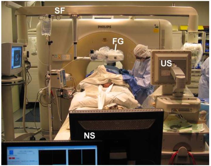
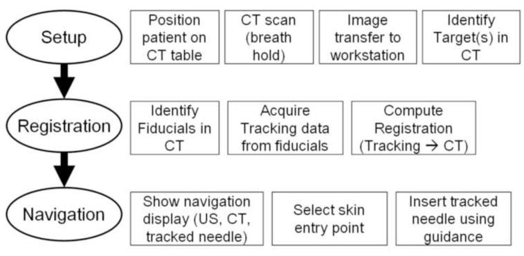
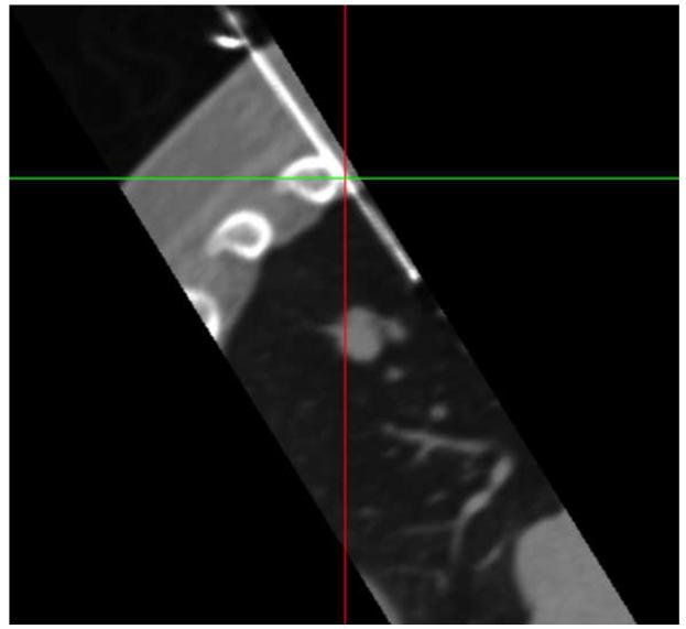
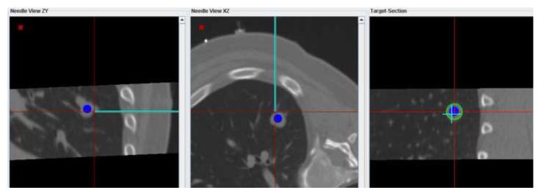
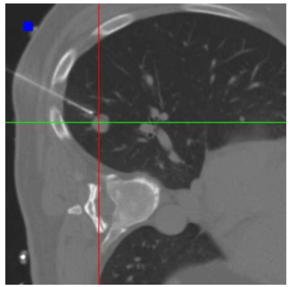
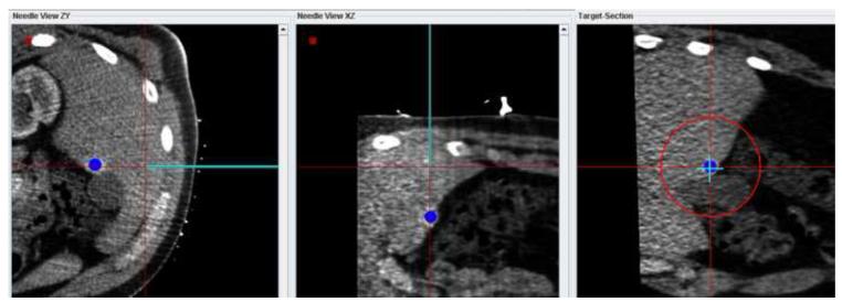
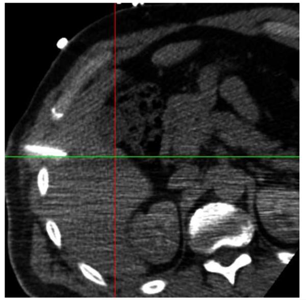
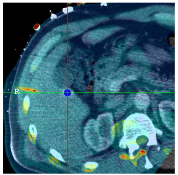
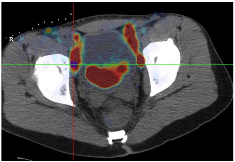
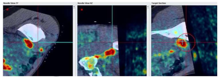
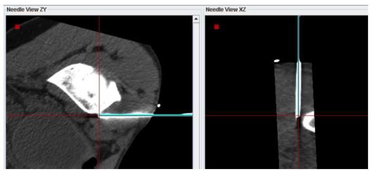
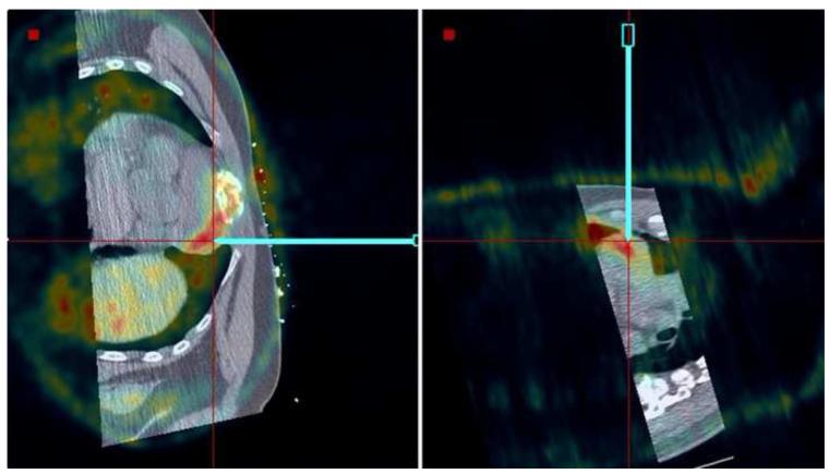
Materials and methods: A combination of conventional image guidance (ultrasound[US]/computed tomography [CT]) and a research navigation system was used in 40 patients undergoing biopsy or RF ablation to assist in target localization and needle and electrode placement. The navigation system displays electromagnetically tracked needles and US images relative to a preprocedural CT scan. Additional images (prior positron emission tomography [PET] or magnetic resonance [MR] imaging) can be fused with CT as needed. Needle aiming with and without tracking were compared, the utility of navigation for each procedure was assessed, the system's off-target tracking error for two different registration methods was evaluated, and setup time was recorded.

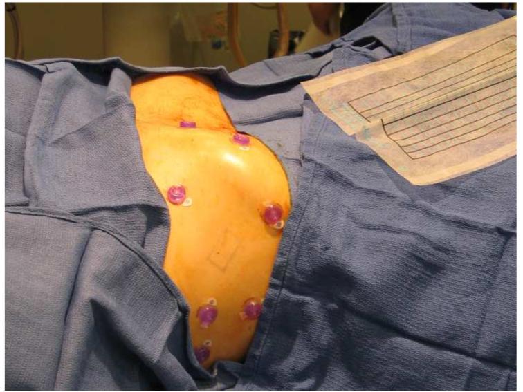
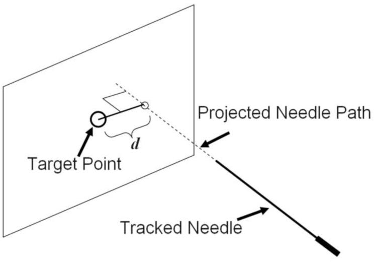
Results: The tracking error could be evaluated in 35 of 40 patients. A basic tracking error of 3.8 mm ± 2.3 was shown using skin fiducial markers for registration. The error improved to 2.7 mm ± 1.6 when using prior internal needle positions as additional fiducial markers. Real-time fusion of US with CT and registration with prior PET and MR imaging were successful and provided clinically relevant guidance information, enabling 19 of the 40 procedures.

Conclusions: The spatial accuracy of the navigation system is sufficient to display clinically relevant image guidance information during biopsy and RF ablation. Breath holding and respiratory gating are effective in minimizing the error associated with tissue motion. In 48% of cases, the navigation system provided information crucial for successful execution of the procedure. Fusion of real-time US with CT or prior diagnostic images may enable procedures that are not feasible with standard, single-modality image guidance.

Trial registration: ClinicalTrials.gov NCT00102544.


Figures

Figure 1
Figure 1
Setup for an US/CT guided biopsy procedure with electromagnetic tracking guidance. FG = electromagnetic field generator, US = Ultrasound scanner, NS = Navigation system workstation, SF = support frame
Figure 2
Figure 2
Procedure workflow using navigation system
Figure 3
Figure 3
Six or seven fiducials were placed on the patient’s skin to facilitate the registration process.
Figure 4
Figure 4
Needle aiming accuracy was determined for conventional guidance and for guidance using the navigation system by calculating the distance d between the target point and the projected needle path at the time of aiming.
Figure 5
Figure 5
(a) MPR showing the biopsy needle and target lesion after 3 unsuccessful attempts to position the needle with CT guidance only (no tracking). (b) Navigation system display showing 3 orthogonal MPRs that were used to bring the virtual needle (cyan) into alignment with the target lesion. (c) Confirmation scan after repositioning the needle with the aid of the navigation system shows the needle well positioned for biopsy.
Figure 5
Figure 5
(a) MPR showing the biopsy needle and target lesion after 3 unsuccessful attempts to position the needle with CT guidance only (no tracking). (b) Navigation system display showing 3 orthogonal MPRs that were used to bring the virtual needle (cyan) into alignment with the target lesion. (c) Confirmation scan after repositioning the needle with the aid of the navigation system shows the needle well positioned for biopsy.
Figure 6
Figure 6
(a) Navigation display showing the virtual needle aligned with the target identified in the arterial phase-enhanced CT. (b) Non-contrast enhanced confirmation scan of the needle position does not show the target lesion. (c) After rigid registration of the confirmation scan (pseudo-colored) with the arterial contrast-enhanced scan (gray scale background) in the vicinity of the target lesion, the correct alignment of the needle with the target is confirmed.
Figure 6
Figure 6
(a) Navigation display showing the virtual needle aligned with the target identified in the arterial phase-enhanced CT. (b) Non-contrast enhanced confirmation scan of the needle position does not show the target lesion. (c) After rigid registration of the confirmation scan (pseudo-colored) with the arterial contrast-enhanced scan (gray scale background) in the vicinity of the target lesion, the correct alignment of the needle with the target is confirmed.
Figure 7
Figure 7
(a) Target selected based on a prior PET scan registered interactively onto the navigation CT scan. (b) The navigation system was used to guide the needle to the hot spot on PET. (c) A verification CT scan showing the actual needle position (white line) was registered with and superimposed on the navigation scan. The two orthogonal MPRs confirm that the virtual needle (blue line) is correctly registered with the image.
Figure 7
Figure 7
(a) Target selected based on a prior PET scan registered interactively onto the navigation CT scan. (b) The navigation system was used to guide the needle to the hot spot on PET. (c) A verification CT scan showing the actual needle position (white line) was registered with and superimposed on the navigation scan. The two orthogonal MPRs confirm that the virtual needle (blue line) is correctly registered with the image.
Figure 8
Figure 8
Needle positioning using navigation relative to a prior PET scan registered and fused with the pre-procedural CT. The target location, a lymphoma directly adjacent to the heart, was clearly visible in the PET scan but was occult in CT.

References

    1. Goldberg SN, Dupuy DE. Image-guided radiofrequency tumor ablation: challenges and opportunities--part I. J Vasc Interv Radiol. 2001;12:1021–1032. - PubMed
    1. Goldberg SN. Radiofrequency tumor ablation: principles and techniques. Eur J Ultrasound. 2001;13:129–147. - PubMed
    1. Dupuy DE, Goldberg SN. Image-guided radiofrequency tumor ablation: challenges and opportunities--part II. J Vasc Interv Radiol. 2001;12:1135–1148. - PubMed
    1. Boss A, Clasen S, Kuczyk M, et al. Magnetic resonance-guided percutaneous radiofrequency ablation of renal cell carcinomas: a pilot clinical study. Invest Radiol. 2005;40:583–590. - PubMed
    1. Krücker J, Xu S, Glossop N, et al. Electromagnetic tracking for thermal ablation and biopsy guidance: clinical evaluation of spatial accuracy. J Vasc Interv Radiol. 2007;18:1141–1150. - PMC - PubMed

Publication types

MeSH terms

Associated data