. 2011 Dec;108(8):1463-75.
doi: 10.1093/aob/mcr033. Epub 2011 Feb 25.

A temporary immersion system improves in vitro regeneration of peach palm through secondary somatic embryogenesis

Affiliations

A temporary immersion system improves in vitro regeneration of peach palm through secondary somatic embryogenesis

D A Steinmacher et al. Ann Bot. 2011 Dec.

Abstract

Background and aims: Secondary somatic embryogenesis has been postulated to occur during induction of peach palm somatic embryogenesis. In the present study this morphogenetic pathway is described and a protocol for the establishment of cycling cultures using a temporary immersion system (TIS) is presented.

Methods: Zygotic embryos were used as explants, and induction of somatic embryogenesis and plantlet growth were compared in TIS and solid culture medium. Light microscopy, scanning electron microscopy (SEM) and transmission electron microscopy (TEM) were used to describe in vitro morphogenesis and accompany morpho-histological alterations during culture.

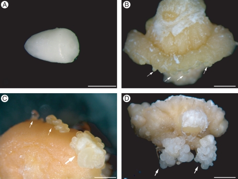
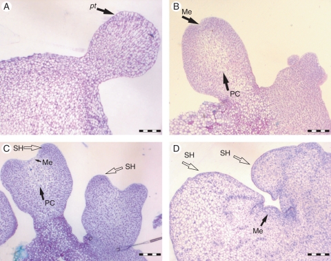
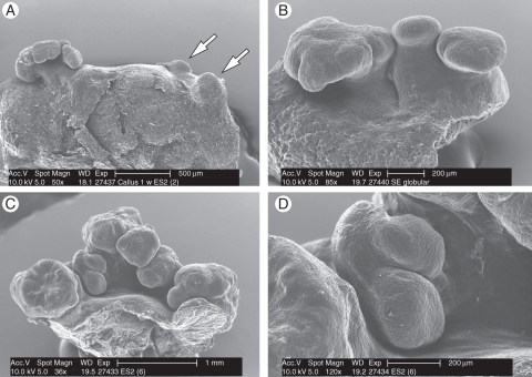
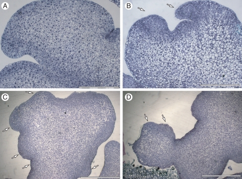
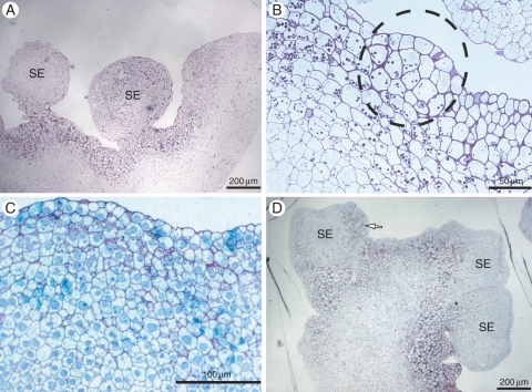
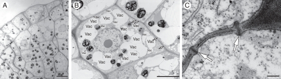
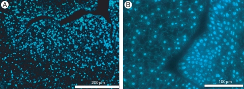
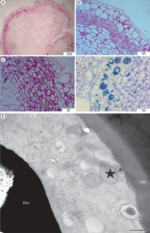
Key results: The development of secondary somatic embryos occurs early during the induction of primary somatic embryos. Secondary somatic embryos were observed to develop continually in culture, resulting in non-synchronized development of these somatic embryos. Using these somatic embryos as explants allowed development of cycling cultures. Somatic embryos had high embryogenic potential (65·8 ± 3·0 to 86·2 ± 5·0 %) over the period tested. The use of a TIS greatly improved the number of somatic embryos obtained, as well as subsequent plantlet growth. Histological analyses showed that starch accumulation precedes the development of somatic embryos, and that these cells presented high nucleus/cytoplasm ratios and high mitotic indices, as evidenced by DAPI staining. Morphological and SEM observations revealed clusters of somatic embryos on one part of the explants, while other parts grew further, resulting in callus tissue. A multicellular origin of the secondary somatic embryos is hypothesized. Cells in the vicinity of callus accumulated large amounts of phenolic substances in their vacuoles. TEM revealed that these cells are metabolically very active, with the presence of numerous mitochondria and Golgi apparatuses. Light microscopy and TEM of the embryogenic sector revealed cells with numerous amyloplasts, large nuclei and nucleoli, and numerous plasmodesmata. Plantlets were obtained and after 3 months in culture their growth was significantly better in TIS than on solid culture medium. However, during acclimatization the survival rate of TIS-grown plantlets was lower.

Conclusions: The present study confirms the occurrence of secondary somatic embryos in peach palm and describes a feasible protocol for regeneration of peach palm in vitro. Further optimizations include the use of explants obtained from adult palms and improvement of somatic embryo conversion rates.


Figures

Fig. 1.
Fig. 1.
Induction of somatic embryogenesis from peach palm zygotic embryos. (A) Mature zygotic embryo of peach palm used as explant. (B) Initial development of globular structures resembling somatic embryos on the callus (arrows) after 4–6 weeks of culture. (C) Further development of globular somatic embryos (arrows) where somatic embryos had previously developed. (D) Development of a cluster of somatic embryos (arrows) on the callus after 6 weeks of culture. Scale bars: (A) = 1 mm, (B) = 3 mm, (C, D) = 2 mm.
Fig. 2.
Fig. 2.
Histological analyses of the development of peach palm somatic embryogenesis. (A) Globular somatic embryo with well-developed protodermis (pt). (B) Elongated somatic embryos showing the initial differentiation of the procambium (PC) and shoot meristem (Me). (C) Mature somatic embryo revealing complete development of the procambium as well as the sheath base (SH). (D) Somatic embryo in conversion conditions revealing a well-formed shoot meristem enclosed by the sheath base. Scale bars = 200 µm.
Fig. 3.
Fig. 3.
Scanning electron microscopy analyses during the induction of peach palm somatic embryos. (A) Initial development of isolated globular structures (arrows). (B) Small clusters of primary somatic embryos. (C) Development of secondary somatic embryos, resulting in a cluster of somatic embryos. (D) Globular somatic embryo revealing the development of secondary somatic embryos. Scale bars: (A) = 500 µm, (C) = 1 mm, (B, D) = 200 µm.
Fig. 4.
Fig. 4.
Histological analyses of the development of peach palm somatic embryos stained with Touludine blue. (A) Histological analysis of peach palm primary somatic embryos revealing intense cell division on sub-epidermal cells. (B) Developed somatic embryo showing intense staining in the sheath base region (arrows). (C) Development of secondary embryos (arrows) from elongated somatic embryo. (D) Initial development of secondary somatic embryos from globular somatic embryos (arrows). Scale bars: (A, B) = 200 µm, (C, D) = 500 µm.
Fig. 5.
Fig. 5.
Histochemical analyses during the development of peach palm secondary somatic embryos (SE). (A) Cluster of somatic embryos from TIS showing high starch accumulation. (B) Possible origin of somatic embryos involving sub-epidermal and epidermal cells (circle). (C) Samples cultured only on solid culture medium. (D) Further development of somatic embryos showing specific starch accumulation in those sectors where other somatic embryos would develop (arrow). Scale bars: (A) = 200 µm, (B) =50 µm, (C) = 100 µm, (D) = 200 µm.
Fig. 6.
Fig. 6.
Ultrastructural analyses of peach palm embryogenic cells. (A) Embryogenic sector including epidermal and sub-epidermal cells. (B) Example of a cell of the embryogenic sector contained numerous small vacuoles (Vac) and numerous plasmodesma (arrows). (C) Aspect of the plasmodesma. Scale bars: (A) = 10 µm, (B) = 5 µm, (C) = 200 nm.
Fig. 7.
Fig. 7.
Mitotic events in the embryogenic sector revealed by DAPI staining. (A) Higher nucleus/cytoplasm ratio observed in sub-epidermal cells. (B) Mitotic events during the initial development of peach palm secondary somatic embryos. Scale bars: (A) = 200 µm, (B) = 100 µm.
Fig. 8.
Fig. 8.
Histological and ultrastructural aspects of the callus sector. (A) General view of callus stained with PAS reagent. (B) Detailed view of A showing an epidermis-like cell layer (arrow). (C) Presence of amyloplast in the callus sector (black arrow). (D) Histological section of the callus sector in contact with the embryogenic sector revealing the accumulation of phenolic substances in the cells. (E) Detailed view of the phenol-storing cells showing numerous mitochondria (stars), and Golgi complex (arrow). Scale bars: (A) = 200 µm, (B, C) = 50 µm, (D) = 500 µm, (E) = 500 nm.

References

    1. Albarran J, Bertrand B, Lartaud M, Etienne H. Cycle characteristics in a temporary immersion bioreactor affect regeneration, morphology, water and mineral status of coffee (Coffea arabica) somatic embryos. Plant Cell Tissue and Organ Culture. 2005;81:27–36.
    1. Alemanno L, Devic M, Niemenak N, et al. Characterization of leafy cotyledon1-like during embryogenesis in Theobroma cacao L. Planta. 2008;227:853–866. - PubMed
    1. Arias O. Propagación vegetativa por cultivo de tejidos del pejibaye (Bactris gasipaes H.B.K.) Asbana. 1985;24:24–27.
    1. Arias O, Huete F. Vegetative propagation in vitro of pejibaye (Bactris gasipaes HBK) Turrialba. 1983;33:103–108.
    1. Bovi MLA, Martins CC, Spiering SH. Desidratação de sementes de quatro lotes de pupunheira: efeitos sobre a germinação e o vigor. Horticultura Brasileira. 2004;22:109–112.

Publication types