. 2011 Jul;39(12):4961-74.
doi: 10.1093/nar/gkr085. Epub 2011 Feb 25.

Sam68 marks the transcriptionally active stages of spermatogenesis and modulates alternative splicing in male germ cells

Affiliations

Sam68 marks the transcriptionally active stages of spermatogenesis and modulates alternative splicing in male germ cells

Maria Paola Paronetto et al. Nucleic Acids Res. 2011 Jul.

Abstract

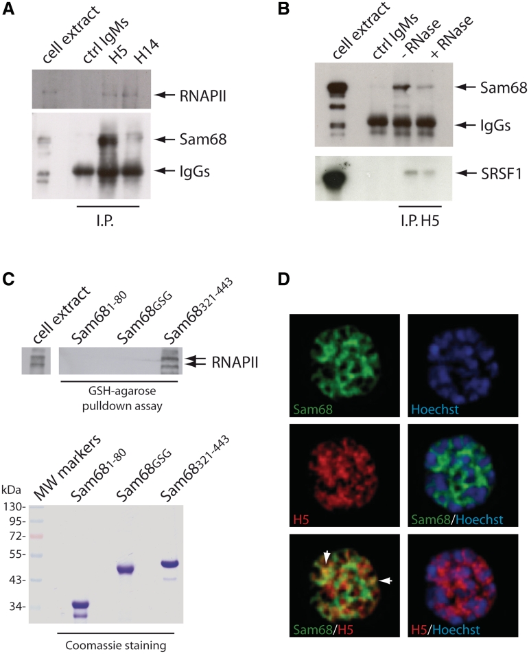
Sam68 plays an essential role in mouse spermatogenesis and male fertility. Herein, we report an interaction between Sam68 and the phosphorylated forms of the RNA polymerase II (RNAPII) in meiotic spermatocytes. RNase treatment decreased but did not abolish the interaction, consistently with in vitro binding of RNAPII to the Sam68 carboxyl-terminal region. Sam68 retention in the spermatocyte nucleus was dependent on the integrity of cellular RNAs, suggesting that the protein is recruited to transcriptionally active chromatin. Mouse knockout models characterized by stage-specific arrest of spermatogenesis and staining with the phosphorylated form of RNAPII documented that Sam68 expression is confined to the transcriptionally active stages of spermatogenesis. Furthermore, Sam68 associates with splicing regulators in germ cells and we report that alternative splicing of Sgce exon 8 is regulated in a Sam68-dependent manner during spermatogenesis. RNA and chromatin crosslink immunoprecipitation experiments showed that Sam68 binds in vivo to sequences surrounding the intron 7/exon 8 boundary, thereby affecting the recruitment of the phosphorylated RNAPII and of the general splicing factor U2AF65. These results suggest that Sam68 regulates alternative splicing at transcriptionally active sites in differentiating germ cells and provide new insights into the regulation of Sam68 expression during spermatogenesis.


Figures

Figure 1.
Figure 1.
Sam68 associates with phosphorylated RNAPII. (A) Co-immunoprecipitation of RNAPII and Sam68. Nuclear extracts from wild-type pachytene spermatocytes (500 µg) were immunoprecipitated with 1 µg of control IgMs, H5 antibody or H14 antibody. Immunoprecipitated proteins were analysed in western blot using anti-RNAPII and anti-Sam68 antibodies as indicated. (B) The co-immunoprecipitation experiment described in (A) was performed using either control IgMs or the H5 antibody and spermatocyte nuclear extracts in the presence or absence of RNase A in the immunoprecipitation buffer. Immunoprecipitated samples were analysed by western blot with anti-Sam68 and anti-SRSF1 antibodies. (C) The western blot analysis of RNAPII in pull-down assay using GST fusion proteins containing the N-terminus (amino acids 1–80), GSG domain or C-terminus (amino acids 321–443) of Sam68 and spermatocyte nuclear extracts. Coomassie staining of the purified GST-fusion proteins used in the assay is shown below. (D) Deconvolution images of a spermatocyte nucleus stained with antibodies for Sam68 (green), serine 2-phosphorylated RNAPII (H5, red). DNA was visualized by Hoechst staining. Merged images are shown as indicated. White arrows indicate overlapping signal of Sam68 and RNAPII in euchromatic regions of the nucleus.
Figure 2.
Figure 2.
Nuclear localization of Sam68 requires the integrity of nucleic acids. Purified pachytene spermatocytes were permeabilized on microscope slides in a buffer containing 0.1% Triton X-100 and incubated for 15 min with medium alone (Control) or DNase or Rnase as indicated. At the end of the incubation, cells were washed three times with PBS and fixed for immunofluorescence analysis with the anti-Sam68 and H5 (A) or H14 (B) antibodies. DNA was stained by Hoechst dye. (C) The western blot analysis of Sam68 and β-tubulin in control or treated (DNase or RNase) pachytene spermatocytes after sequential extractions with the indicated buffers.
Figure 3.
Figure 3.
Stage-specific expression of Sam68 during meiotic prophase. (A) Immunohistochemical analysis of Sam68 expression in adult mouse testis. Mouse testicular sections representing the indicated stages of the cycle of the seminiferous tubule were stained with anti-Sam68 antibody and counterstained with hematoxilin to detect cell nuclei. Arrows indicate representative pachytene spermatocytes with different expression levels of the protein. The stage of the seminiferous tubule was assessed by staining an adjacent section with PAS and it is labelled in the lower left side of each panel. (B) Co-staining of testicular sections from adult wild-type mice with anti-Sam68 (red) and anti-H1t (green) antibodies. Nuclei were visualized by DAPI staining. Representative pachytene (Pc) spermatocytes are indicated by arrows. (C) Co-staining of nuclear spreads obtained from mouse spermatocytes with antibodies against SCP-3 (red) and Sam68 (green). DAPI was used for the DNA staining (blue). Sam68 is absent in leptotene (ad), zygotene (eh) and early pachytene spermatocytes (i–l, early Pc indicated by the arrow), but it is expressed in mid pachytene (m–p) and diplotene (i–l) spermatocytes.
Figure 4.
Figure 4.
Sam68 expression in knockout models of meiotic arrest. (A) Sam68 staining (red) in Atm/ (a–d), H2AX/ (e–h) and Mlh/ testis (i–l). Zg, zygotene; Pc, pachytene; St, Sertoli cells. Sam68 is strongly expressed by the somatic Sertoli cells at the basis of the tubules in the Atm−/− and H2ax−/− testis, where meiosis is arrested at the zygotene/early pachytene stage. In Mlh1−/− mice, spermatocytes progress through prophase I and Sam68 was highly expressed in the meiotic cells (i–l). Panels d, h and l are enlarged images of c, g and k. (B) Sam68 staining (red) in Atm−/−Spo11+/ (a–c) and Spo11−/− testis (d–f). Sam68 is expressed in Pc spermatocyes of Atm−/−Spo11+/ mice and in Pc-like spermatocytes of Spo11−/− mice, respectively.
Figure 5.
Figure 5.
Sam68 is co-expressed with phosphorylated RNAPII in male germ cells. (A) Testis section from a 25-day-old wild-type mouse was stained with anti-Sam68 (a) and H5 (b; anti-pser2 RNAPII) antibodies and co-stained with Hoechst (c) to detect nuclei. Arrows indicate the pachytene (Pc) and zygotene (Zg) spermatocytes. (B) Wild-type male germ cells were isolated by elutriation and analysed by immunofluorescence using the anti-Sam68 (a,e) and H5 (b,f; anti-p-ser2 RNAPII) antibodies as indicated. Nuclei were stained with Hoechst (c,g) to identify cell stages by nuclear morphology. Arrows in panels d and h indicate the zygotene (Zg) and pachytene (Pc) spermatocytes in the population examined. (C) Immunofluorescence analysis of isolated germ cells with the anti-Sam68 (a) and anti-γH2AX (b) antibodies. Nuclei were stained with Hoechst (c). In leptotene spermatocytes (Lp; arrows in panel d), γH2AX is localized in nuclear foci and Sam68 is absent; in pachytene spermatocytes (Pc; arrows in panel d), γH2AX is localized in the sex body whereas Sam68 is excluded from it.
Figure 6.
Figure 6.
Sam68 regulates alternative splicing of Sgce exon 8 in male germ cells. RT–PCR analysis of Sgce exon 8 inclusion in total RNA extracted from Sam68 wild-type or knockout testes at 8, 16 and 30 days post partum (dpp) (A) or from isolated wild-type or knockout spermatocytes and spermatids. (B) Bands corresponding to mRNAs containing or not, exon 8 are indicated on the right of the panels. (C and D) Chromatin immunoprecipitation (ChIP) analysis of Sam68 wild-type and knockout germ cells. Sonicated chromatin (100 µg of DNA/sample) was immunoprecipitated with 2µg of H5, H14 antibodies or control rabbit IgGs and co-precipitated DNA was analysed by real time PCR with primers (black arrows in the scheme) spanning the Sgce transcription unit as indicated. (E) CLIP analysis of the binding of Sam68 to Sgce pre-mRNA. After UV crosslink, Sam68 wild-type and knockout germ cell extracts were sonicated and treated with DNase and RNase to yield RNA fragments of ∼200 to 250 nt. Wild-type and knockout germ cell extracts were immunoprecipitated with 2 µg of rabbit IgGs or anti-Sam68 antibodies and co-precipitated RNA was analysed by real time PCR with primers (black arrows in the scheme) spanning the Sgce transcription unit as indicated. (F) Electrophoretic mobility shift assays (EMSAs) of the binding of purified GST-Sam681–277 to a labelled Sgce probe containing the sequences encoded at the intron 7/exon 8 boundary. The position of the free probe and the probe complexed with GST-Sam681–277 are shown on the left side. Competition with the wild-type (middle panel) or mutated (right panel) cold probes is shown. The scheme above the gels shows the wild-type and mutated sequence used in the EMSAs. The mutated bases that interfere with the Sam68 consensus are shown in violet. (G) The western blot analysis of RNA pulldown assay of U2AF65 binding to Sgce exon 8. Biotinylated RNAs encoding the Sgce wild-type and mutated exon 8 sequences (from −32 to +63) were bound to Streptavidine agarose beads and incubated with testicular nuclear extract (100 µg) in the presence of 1 µg of purified GST or GST-Sam681–277, as indicated.

Similar articles

Cited by

References

    1. Braun RE. Post-transcriptional control of gene expression during spermatogenesis. Semin. Cell Dev. Biol. 1998;9:483–489. - PubMed
    1. Elliott D. Pathways of post-transcriptional gene regulation in mammalian germ cell development. Cytogenet. Genome Res. 2003;103:210–216. - PubMed
    1. Paronetto MP, Sette C. Role of RNA-binding proteins in mammalian spermatogenesis. Int. J. Androl. 2010;33:2–12. - PubMed
    1. Monesi V. Ribonucleic acid synthesis during mitosis and meiosis in the mouse testis. J. Cell Biol. 1964;22:521–532. - PMC - PubMed
    1. Turner JM, Mahadevaiah SK, Fernandez-Capetillo O, Nussenzweig A, Xu X, Deng CX, Burgoyne PS. Silencing of unsynapsed meiotic chromosomes in the mouse. Nat. Genet. 2005;37:41–47. - PubMed

Publication types