. 2011 Mar;22(3):460-71.
doi: 10.1681/ASN.2010060681. Epub 2011 Feb 25.

Activation of PI3-kinase stimulates endocytosis of ROMK via Akt1/SGK1-dependent phosphorylation of WNK1

Affiliations

Activation of PI3-kinase stimulates endocytosis of ROMK via Akt1/SGK1-dependent phosphorylation of WNK1

Chih-Jen Cheng et al. J Am Soc Nephrol. 2011 Mar.

Abstract

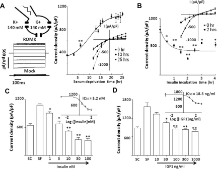
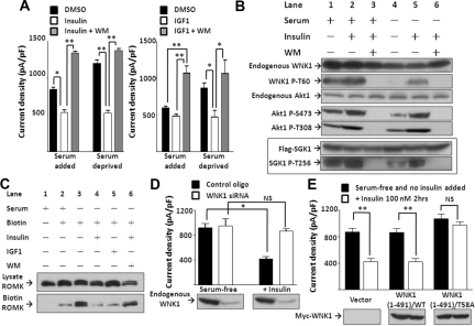
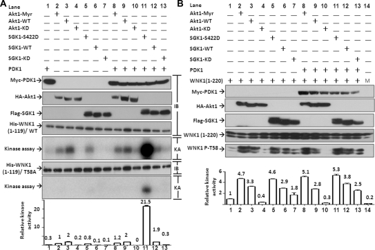
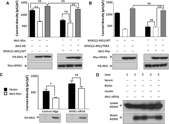
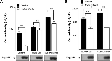
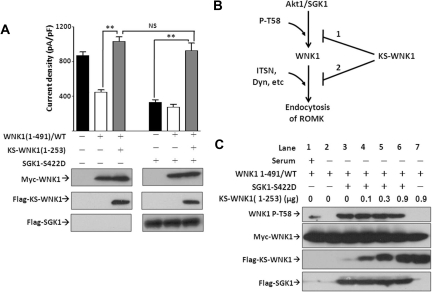
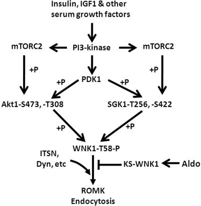
WNK kinases stimulate endocytosis of ROMK channels to regulate renal K+ handling. Phosphatidylinositol 3-kinase (PI3K)-activating hormones, such as insulin and IGF 1, phosphorylate WNK1, but how this affects the regulation of ROMK abundance is unknown. Here, serum starvation of ROMK-transfected HEK cells led to an increase of ROMK current density; subsequent addition of insulin or IGF1 inhibited ROMK currents in a PI3K-dependent manner. Serum and insulin also increased phosphorylation of the downstream kinases Akt1 and SGK1 as well as WNK1. A biotinylation assay suggested that insulin and IGF1 inhibit ROMK by enhancing its endocytosis, a process that WNK1 may mediate. Knockdown of WNK1 with siRNA or expression of a phospho-deficient WNK1 mutant (T58A) both prevented insulin-induced inhibition of ROMK currents, suggesting that phosphorylation at Threonine-58 of WNK1 is important to mediate the inhibition of ROMK by PI3K-activating hormones or growth factors. In vitro and in vivo kinase assays supported the notion that Akt1 and SGK1 can phosphorylate WNK1 at this site, and we established that Akt1 and SGK1 synergistically inhibit ROMK through WNK1. We used dominant-negative intersectin and dynamin constructs to show that SGK1-mediated phosphorylation of WNK1 inhibits ROMK by promoting its endocytosis. Taken together, these results suggest that PI3K-activating hormones inhibit ROMK by enhancing its endocytosis via a mechanism that involves phosphorylation of WNK1 by Akt1 and SGK1.


Figures

Figure 1.
Figure 1.
Insulin and IGF1 inhibit ROMK current. (A) Effect of serum deprivation on ROMK current. (Left) Configuration of whole-cell recording, voltage-clamp protocol (from −100 to 100 mV), representative currents from ROMK- and mock-transfected cells are shown. (Right) ROMK current density (pA/pF at −100 mV; normalized to the cell surface area) at different time of serum deprivation are shown (mean ± SEM, n ≥ 6 for each) and analyzed by nonlinear regression curve. Inset shows current-voltage (I-V) relationship curve of ROMK with serum deprivation for 0, 13, and 25 hours. Data in each time point was compared with serum-containing group (0-hour serum deprivation). **P < 0.01. All time points beyond 6 hours are significant compared with the serum-containing group (not indicated by asterisk). All time points between 16 and 25 hours are not significantly different (not indicated). In all experiments throughout this study, ROMK currents shown are after subtracting residual currents in the presence of 5 mM barium. (B) Time course of effect of insulin on serum-deprived ROMK current. Cells were cultured in serum-free medium at least 16 hours before addition of insulin (100 nM) for different time periods. Data points are mean ± SEM (n ≥ 6 for each), compared with serum-deprived (0-hour insulin incubation), and analyzed by nonlinear regression curve. Inset shows I-V curve of ROMK current before and after 2-hour insulin. (C and D) Dose–response curve of insulin and IGF1 on serum-deprived ROMK. ROMK current density (mean ± SE, n ≥ 6) at −100 mV was measured in cells cultured in serum-containing medium (SC), serum-free medium (SF), and 2-hour incubation of different concentration of insulin or IGF1. Data of each insulin or IGF1 treatment group were compared with the SF group. Dose–response curve and IC50 of insulin or IGF1 on ROMK resulted from nonlinear regression analysis. *P < 0.05 versus designated group by unpaired two-tailed t test. **P < 0.01.
Figure 2.
Figure 2.
Insulin and IGF1 inhibit ROMK through PI3K and WNK1–T58 phosphorylation. (A) Effect of insulin and IGF1 on ROMK is blocked by wortmannin. Cells cultured with or without serum were treated by DMSO, insulin 100 nM, or insulin 100 nM plus wortmannin (WM, 100 nM), respectively, for 2 hours (mean ± SEM, n ≥ 6). (B) Effect of serum, insulin, and wortmannin on phosphorylation of endogenous WNK1, Akt1, and overexpressed SGK1. Phosphorylation on specific residues was determined by specific anti-phospho antibodies. Shown is representative of three separate experiments of similar results. (C) Effect of insulin and IGF1 on surface abundance of ROMK. Cells were serum-deprived for 16 hours and treated with insulin (100 nM), IGF1 (100 ng/ml), and/or WM (100 nM) for 2 hours as indicated before biotinylation (Biotin). ROMK in total cell lysates (Lysate ROMK) and elute from avidin beads (Biotin ROMK) were detected by Western blot. (D) Insulin inhibits ROMK through WNK1. Cells transfected with control oligonucleotide or WNK1 siRNA (200 nM each) were deprived of serum for 16 hours (serum-free group). Insulin (100 nM) was added for 2 hours (+Insulin group). Successful knockdown of endogenous WNK1 by siRNA is evident by Western blot analysis. Mean ± SEM (n ≥ 6 each). (E) Insulin inhibits ROMK through phosphorylation on T58 of WNK1. Cells transfected with empty vector, wild-type (WT), or T58A mutant WNK1(1-491) were cultured in serum-free medium for 16 hours and received insulin (100 nM) or not for 2 hours. Equal amount of WT or T58A WNK1(1-491) expression is evident by Western blot analysis. Mean ± SEM (n ≥ 6 each). In A, D, and E, *P < 0.05 between indicated groups by unpaired two-tailed t test. **P < 0.01. NS, not statistically significant.
Figure 3.
Figure 3.
Akt1 and SGK1 phosphorylate WNK1 at threonine-58. (A) In vitro kinase assay of PDK1, Akt1, and SGK1. Epitope-tagged (Myc-, HA-, or Flag-) wild-type SGK1 (SGK1-WT), kinase-dead SGK1 (SGK1-KD), constitutively active mutant SGK1 (SGK1–S422D), WT Akt1 (Akt1-WT), myristoylated-Akt1 (Akt1-Myr), and kinase-dead Akt1 (Akt1-KD) were expressed in HEK cells with or without wild-type PDK1. Kinase activity of immunoprecipitated PDK1, Akt1, and SGK1 was assayed using wild-type or T58A WNK1(1-119) as a substrate. Immunoblots of precipitated proteins by respective epitope antibody (labeled “IB” on the right) and autoradiograph of kinase assay analyzed by phosphoimager (labeled “KA” on the right) are shown. Bar graph in the bottom is the relative kinase activity (normalized to lane 2, “Akt1-Myr”) specific for phosphorylation at T58 [i.e., subtracting non-T58 phosphorylation signal on WNK1(1-119)/T58A mutant from signal on wild-type WNK1(1-119)]. Mean ± SEM from three separate experiments is shown on top of each bar. (B) In vivo phosphorylation on T58 of WNK1 by Akt1 and SGK1. Cells were transfected with epitope-tagged Akt1, SGK1, or PDK1 with wild-type WNK1(1-220) (lanes 1 to 13) or with WNK1(1-220)/T58A mutant (lane 14 labeled “M”). Protein expressions were blotted by specific antibodies. Phosphorylation on T58 of WNK1(1-220) was detected by anti-phospho-T58 WNK1 antibody. Doublet bands detected by anti-WNK1 and anti-phospho-T58 WNK1 antibodies were always found in WNK1(1-119) and WNK1(1-220) but not WNK1(1-491). The doublets probably represent different conformational forms of eukaryotic WNK1 proteins because they are not observed for purified His-tagged WNK1(1-119) proteins produced in the bacteria (see A). The abundance of each band in the gel reflecting kinase activity of Akt1 or SGK1 was measured by densitometry by the Image J program available at the NIH website. Basal level of T58 phosphorylation on WNK1(1-220) without exogenous Akt1 or SGK1 (lane 1) was defined as 1 for relative kinase activity measurement. Mean ± SEM from three separate experiments is shown on top of each bar.
Figure 4.
Figure 4.
Akt1 inhibits ROMK in a WNK1–T58 phosphorylation-dependent manner. (A) Effect of Akt1 on ROMK. Cells were transfected with ROMK and with myristoylated Akt1 (Akt1-Myr), kinase-dead Akt1 (Akt1-KD), and/or WNK1(1-491) as indicated. (B) Effect of myristoylated Akt1 (Akt1-Myr) on ROMK in the presence of wild-type (WT) or T58A mutant of WNK1(1-491). (C) Effect of Akt1 on ROMK with or without endogenous WNK1. Cells were transfected with control oligonucleotide or WNK1 siRNA (200 nM each) and with empty vector or myristoylated Akt1 (Akt1-Myr). In each panel, ROMK current density (pA/pF at −100 mV) was represented as mean ± SEM (n ≥ 6). *P < 0.05 between indicated groups by unpaired two-tailed t test. **P < 0.01. NS, not statistically significant. Equal protein expression was confirmed by Western blot. (D) Effect of insulin on membrane abundance of ROMK with or without endogenous Akt1. Cells were transfected with ROMK and control oligonucleotide or Akt1 siRNA (200 nM each) and deprived of serum for 16 hours.
Figure 5.
Figure 5.
SGK1 inhibits ROMK through the same pathway as Akt1. (A) Effect of SGK1 on ROMK. Cells were co-transfected with ROMK and constitutively active SGK1 (SGK1–S422D) or kinase-dead SGK1 (SGK1-KD) with or without WNK1(1-491). (B) Effect of SGK1 on ROMK in the presence of wild-type or T58A mutant of WNK1(1-491). SGK1–S422D was co-transfected with wild-type or T58A mutant WNK1(1-491). (C) Effect of SGK1 on ROMK with or without endogenous WNK1. Cells were transfected with control oligonucleotide or WNK1 siRNA (200 nM each) and with empty vector or SGK1–S422D. (D) Effect of siRNA of Akt1 and/or SGK1 on ROMK. Cells were transfected with siRNA for Akt1 (siAkt1) and/or SGK1 (siSGK1, 200 nM each) and with vector or WNK1(1-491). In each panel, ROMK current density was measured and presented as mean ± SEM (n ≥ 6 for each group). *P < 0.05 between indicated groups by unpaired two-tailed t test. **P < 0.01. NS, not statistically significant. Efficacy of Akt1 siRNA and equal expression of protein were confirmed by Western blot.
Figure 6.
Figure 6.
Effect of SGK1 on ROMK is dynamin and intersectin (ITSN)-dependent, but not dependent on phosphorylation of ROMK at S44. (A) Effect of SGK1 on ROMK in the presence of dominant-negative intersectin (ITSN-DN) or dynamin (dynamin-DN). Cells were transfected with SGK1–S422D or without (“vector”) and with ITSN-DN or dynamin-DN or without (“control”). (B) Effect of SGK1 on wild-type or S44D mutant ROMK. Cells were co-transfected with SGK1–S422D and with wild-type ROMK (ROMK-WT) or S44D mutant of ROMK (ROMK-S44D). In each panel, ROMK current density was measured and presented as mean ± SEM (n ≥ 6 for each group). *P < 0.05 between indicated groups by unpaired two-tailed t test. **P < 0.01. NS, not statistically significant. Equal protein expression was confirmed by Western blot.
Figure 7.
Figure 7.
Effect of SGK1 on ROMK is reversed by kidney-specific WNK1 (KS-WNK1). (A) Effect of SGK1 on ROMK in the presence of KS-WNK1. Cells were transfected with SGK1–S422D, WNK1(1-491), and/or KS-WNK1(1-253) as indicated. KS-WNK1(1-253) consists of amino acids 1 to 253 of KS-WNK1. ROMK current density was measured and presented as mean ± SEM (n ≥ 6 for each group). **P < 0.01 between indicated groups by unpaired two-tailed t test. NS, not statistically significant. Equal protein expression was confirmed by Western blot. (B) Possible mechanisms for KS-WNK1 to block SGK1 effect on ROMK. Mechanism 1: Interfering with WNK1 phosphorylation by SGK1. Mechanism 2: Interfering with WNK1 interaction with downstream effectors, such as intersectin (“ITSN”) and dynamin (“Dyn”). (C) Effect of KS-WNK1 on WNK1–T58 phosphorylation by SGK1. Cells were all transfected with WNK1(1-491), KS-WNK1(1-253) (at DNA amount from 0 to 0.9 μg), and/or SGK1–S422D and incubated with or without serum as indicated. Basal level of WNK1(1-491) phosphorylation is shown in lane 1. For experiments shown in lanes 2 to 7, cells were incubated in serum-free media for 16 hours. Protein expression was detected by specific antibodies. Phosphorylation on WNK1–T58 was determined using anti-phospho-T58 WNK1 antibody.
Figure 8.
Figure 8.
A working model for regulation of ROMK by PI3K-activating hormones via Akt1/SGK1 and WNK1 and by aldosterone. See texts for details and for abbreviations.

Similar articles

Cited by

References

    1. Ho K, Nichols CG, Lederer WJ, Lytton J, Vassilev PM, Kanazirska MV, Hebert SC: Cloning and expression of an inwardly rectifying ATP-regulated potassium channel. Nature 362: 31–38, 1993 - PubMed
    1. Rodan AR, Huang CL: Distal potassium handling based on flow modulation of maxi-K channel activity. Curr Opin Nephrol Hypertens 18: 350–355, 2009 - PMC - PubMed
    1. Zeng WZ, Babich V, Ortega B, Quigley R, White SJ, Welling PA, Huang CL: Evidence for endocytosis of ROMK potassium channel via clathrin-coated vesicles. Am J Physiol Renal Physiol 283: F630–F639, 2002 - PubMed
    1. Xu B, English JM, Wilsbacher JL, Stippec S, Goldsmith EJ, Cobb MH: WNK1, a novel mammalian serine/threonine protein kinase lacking the catalytic lysine in subdomain II. J Biol Chem 275: 16795–16801, 2000 - PubMed
    1. Wilson FH, Disse-Nicode‘me S, Choate KA, Ishikawa K, Nelson-Williams C, Desitter I, Gunel M, Milford DV, Lipkin GW, Achard JM, Feely MP, Dussol B, Berland Y, Unwin RJ, Mayan H, Simon DB, Farfel Z, Jeunemaitre X, Lifton RP: Human hypertension caused by mutations in WNK kinases. Science 293: 1107–1112, 2001 - PubMed

Publication types

MeSH terms

Substances

LinkOut - more resources