

Fibroblasts and myofibroblasts in renal fibrosis
- PMID: 21355940
- PMCID: PMC3101489
- DOI: 10.1111/j.1365-2613.2011.00764.x
Fibroblasts and myofibroblasts in renal fibrosis
Abstract
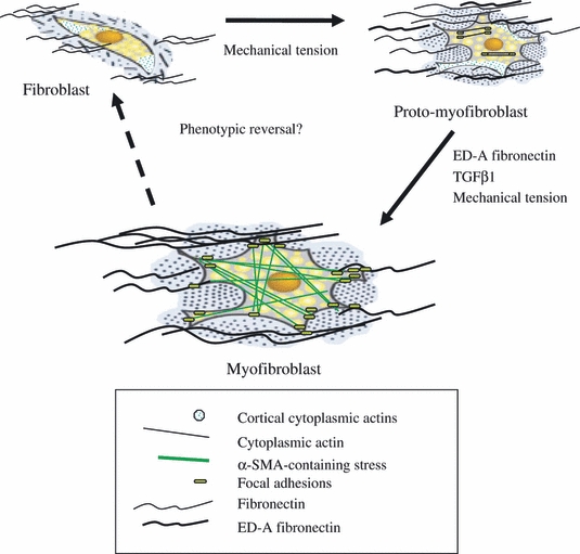
Interstitial fibrosis, associated with extensive accumulation of extracellular matrix constituents in the cortical interstitium, is directly correlated to progression of renal disease. The earliest histological marker of this progression is the accumulation in the interstitium of fibroblasts with the phenotypic appearance of myofibroblasts. These myofibroblasts are contractile cells that express alpha smooth muscle actin and incorporate it into intracellular stress fibres. Although fibroblasts are histologically visible in normal kidneys, there are relatively few of them and proximal tubular epithelial cells predominate. In progressive disease, however, the interstitium becomes filled with myofibroblasts. In this review, we will examine the phenotype and function of fibroblasts and myofibroblasts in the cortical interstitium and the processes that may modulate them.
© 2011 The Authors. International Journal of Experimental Pathology © 2011 International Journal of Experimental Pathology.
Figures
References
-
- Abe R, Donnelly SC, Peng T, Bucala R, Metz CN. Peripheral blood fibrocytes: differentiation pathway and migration to wound sites. J. Immunol. 2001;166:7556–7562. - PubMed
-
- Alpers CE, Hudkins KL, Floege J, Johnson RJ. Human renal cortical interstitial cells with some features of smooth muscle cells participate in tubulointerstitial and crescentic glomerular injury. J. Am. Soc. Nephrol. 1994;5:210–219. - PubMed
-
- Attisano L, Wrana JL. Signal transduction by the TGF-beta superfamily. Science. 2002;296:1646–1647. - PubMed
-
- Broekema M, Harmsen MC, van Luyn MJ, et al. Bone marrow-derived myofibroblasts contribute to the renal interstitial myofibroblast population and produce procollagen I after ischemia/reperfusion in rats. J. Am. Soc. Nephrol. 2007;18:165–175. - PubMed
Publication types
MeSH terms
LinkOut - more resources
Full Text Sources
Other Literature Sources
