. 2011 Aug;25(6):1113-22.
doi: 10.1016/j.bbi.2011.02.008. Epub 2011 Feb 26.

Brain inflammation is induced by co-morbidities and risk factors for stroke

Affiliations

Brain inflammation is induced by co-morbidities and risk factors for stroke

Caroline Drake et al. Brain Behav Immun. 2011 Aug.

Abstract

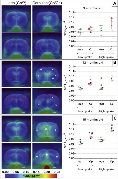
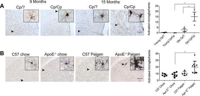
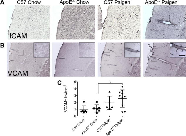
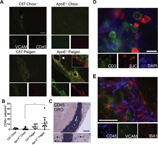
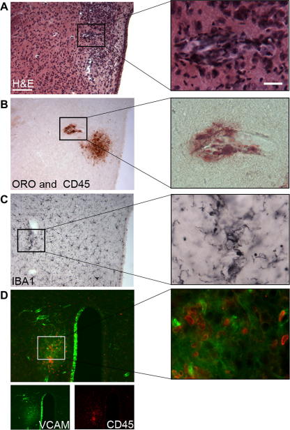
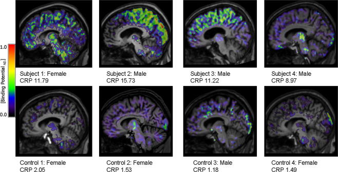
Chronic systemic inflammatory conditions, such as atherosclerosis, diabetes and obesity are associated with increased risk of stroke, which suggests that systemic inflammation may contribute to the development of stroke in humans. The hypothesis that systemic inflammation may induce brain pathology can be tested in animals, and this was the key objective of the present study. First, we assessed inflammatory changes in the brain in rodent models of chronic, systemic inflammation. PET imaging revealed increased microglia activation in the brain of JCR-LA (corpulent) rats, which develop atherosclerosis and obesity, compared to the control lean strain. Immunostaining against Iba1 confirmed reactive microgliosis in these animals. An atherogenic diet in apolipoprotein E knock-out (ApoE(-/-)) mice induced microglial activation in the brain parenchyma within 8 weeks and increased expression of vascular adhesion molecules. Focal lipid deposition and neuroinflammation in periventricular and cortical areas and profound recruitment of activated myeloid phagocytes, T cells and granulocytes into the choroid plexus were also observed. In a small, preliminary study, patients at risk of stroke (multiple risk factors for stroke, with chronically elevated C-reactive protein, but negative MRI for brain pathology) exhibited increased inflammation in the brain, as indicated by PET imaging. These findings show that brain inflammation occurs in animals, and tentatively in humans, harbouring risk factors for stroke associated with elevated systemic inflammation. Thus a "primed" inflammatory environment in the brain may exist in individuals at risk of stroke and this can be adequately recapitulated in appropriate co-morbid animal models.


Figures

Fig. 1
Fig. 1
Sum images (20–60 min post-injection; left panel) and respective quantification (graphs on the right panel) of [18F]DPA-714 uptake in the brain of lean (+/?) and corpulent (cp/cp) rats at 9 (A), 12 (B) and 15 months (C) of age. * and ‡ indicate a significant difference between lean and corpulent animals of the same age in respectively low and high uptake regions of interest (P < 0.05, Mann–Whitney test). # indicates a significant difference between 9 (A) and 15 (B) months old animals (P < 0.05, Mann–Whitney test). Data are expressed as and mean ± SD (filled symbols correspond to the respective image on the left panel).
Fig. 2
Fig. 2
Rodent models of atherosclerosis involve microglial activation in the brain. (A) Activated microglia as identified by increased Iba1 immunopositivity, thickened processes and irregular cell bodies were seen in the striatum of 15 month old corpulent rats, but not in 9 month old animals. Aged corpulent rats had a significantly increased number of activated microglia compared to young corpulent, or 15 month old heterozygous rats. (B) Activated, Iba1-positive microglia was numerous in ApoE−/− mice fed a Paigen diet. Insets show representative images of microglial cells from the different groups of mice. Quantitative analysis revealed significantly more activated microglial cells in the striatum of ApoE−/− mice fed a Paigen diet compared with ApoE−/− mice fed chow diet. P < 0.05. Scale bars: 200 and 10 μm (insets).
Fig. 3
Fig. 3
Cerebrovascular activation occurs in the brain in association with peripheral atherosclerosis. Vascular activation was assessed in the cerebral cortex using immunostaining to the adhesion molecules (A) ICAM and (B) VCAM. Unlike mice fed a chow diet, mice fed a Paigen diet showed an increased number of ICAM and VCAM-positive blood vessels in the brain. (C) Quantitative analysis of VCAM-positive blood vessels in the cerebral cortex. Scale bars: 200 and 50 μm (inset).
Fig. 4
Fig. 4
Microglia/macrophages, granulocytes and T cells accumulate in the choroid plexus of the caudal lateral ventricle in response to peripheral atherosclerosis. (A) ApoE−/− mice fed a Paigen diet show accumulation of CD45+ leucocytes (red) in the choroid plexus of the caudal lateral ventricle, which display increased VCAM (green) immunopositivity. (B) Quantification of CD45+ leucocytes in the choroid plexus of the lateral ventricles. (C) CD45-positive cells, which are numerous in the choroid plexus, also appear in the parenchyma (Oil red O counterstain) on both sides of the lateral ventricle (inset, arrowheads). (D) CD3 positive T cells (green) were found to accumulate in a partially overlapping area with granulocytes, identified with an anti-neutrophil serum (SJC, red). (E) A population of microglia/macrophages (Iba1, red) shows increased CD45 immunopositivity (blue) in the caudal choroid plexus among other CD45-positive leucocytes (possibly granulocytes). P < 0.05. Scale bars: A; 200 μm; C; 100 μm; D; 10 μm and E; 50 μm.
Fig. 5
Fig. 5
Focal pathological changes are present in the brain in response to peripheral atherosclerosis. Haematoxylin & Eosin (H&E) staining (A) reveals vascular inflammation as indicated by dilated blood vessels and inflammatory infiltrates in the hypothalamus adjacent to the third ventricle in Paigen fed ApoE−/− mice. Focal lipid deposition as identified by Oil red O staining is observed in the vicinity of perivascular CD45-positive leucocytes (B). This is associated with an increase in the number of activated, Iba1-positive microglia (C) recruitment of CD45+ cells (D, red) and focally upregulated VCAM immunostaining (D, green). VCAM expression is also seen in the ipsilateral wall of the third ventricle but not in the contralateral part. Parallel brain sections from a representative brain are shown. P < 0.05. Scale bars: 100 and 12.5 μm. (For interpretation of the references to colour in this figure legend, the reader is referred to the web version of this paper.)
Fig. 6
Fig. 6
[11C](R)-PK11195 binding potential (BPND) images are shown for all subjects and control participants. Images are displayed on each subject’s respective T1 MRI scan normalised to the SPM5 T1 brain template. The value for each individual’s CRP at the time of PET scanning is also shown.

References

    1. Allan S.M., Tyrrell P.J., Rothwell N.J. Interleukin-1 and neuronal injury. Nat. Rev. 2005;5:629–640. - PubMed
    1. Amor S., Puentes F., Baker D., van der Valk P. Inflammation in neurodegenerative diseases. Immunology. 2010;129:154–169. - PMC - PubMed
    1. Anthony D., Dempster R., Fearn S., Clements J., Wells G., Perry V.H., Walker K. CXC chemokines generate age-related increases in neutrophil-mediated brain inflammation and blood–brain barrier breakdown. Curr. Biol. 1998;8:923–926. - PubMed
    1. Boellaard R. Standards for PET image acquisition and quantitative data analysis. J. Nucl. Med. 2009;50(Suppl. 1):11S–20S. (Epub 2009 April 20) - PubMed
    1. Boutin, H., Prenant, C., Galea, J., Greenhalgh, A., Julyan, P., Brown, G., Herholz, K., Rothwell, N., Kassiou, M., 2008. [18F]DPA-714: a new ligand for neuroinflammation, evaluation in a model of cerebral ischemia in rats. First World Molecular Imaging Congress, Program No. 0629.

Publication types

MeSH terms