. 2011 Mar;178(3):1167-76.
doi: 10.1016/j.ajpath.2010.11.055.

γδ T cells attenuate bleomycin-induced fibrosis through the production of CXCL10

Affiliations

γδ T cells attenuate bleomycin-induced fibrosis through the production of CXCL10

Derek A Pociask et al. Am J Pathol. 2011 Mar.

Abstract

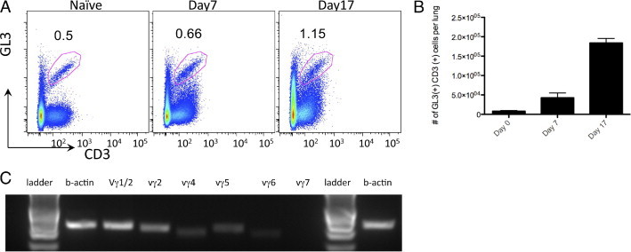
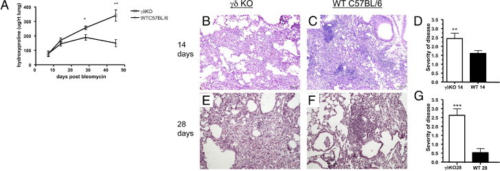
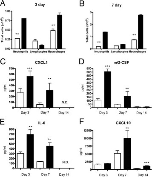
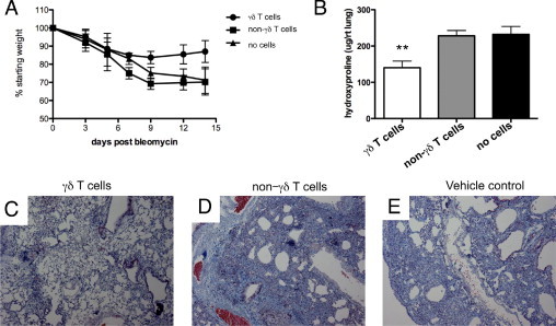
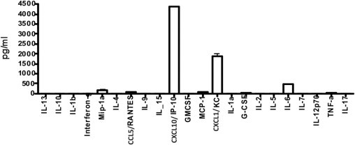
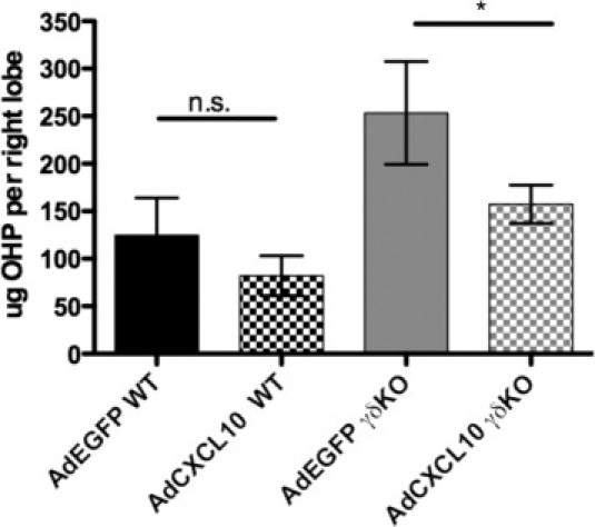
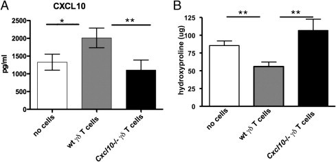
γδ T cells are a subset of T cells associated with epithelial mucosal tissues and play a prominent role in both promoting and dampening inflammatory responses to pathogens; in addition, they strongly mediate epithelial repair. By using a bleomycin model of pulmonary fibrosis, we found that γδ T-cell populations dramatically increased after bleomycin administration. To determine the importance of these cells, we exposed mice lacking the δ chain of the γδ T-cell receptor (γδ knockout [KO]) to bleomycin. Pulmonary fibrosis was more severe in γδ KO mice, as measured by collagen deposition (hydroxyproline) and histopathological features. Furthermore, there was no evidence of resolution of the fibrotic response up to 45 days after bleomycin therapy. In contrast to control mice, γδ KO mice had decreased concentrations of IL-6, granulocyte colony stimulating factor, chemokine CXC ligand (CXCL) 1, and interferon inducible protein 10/CXCL10. In vitro culture of γδ T cells purified from lungs 17 days after bleomycin exposure (a time of peak influx of these cells) demonstrated that γδ T cells produced substantial quantities of all four of these cytokines, suggesting that γδ T cells are a predominant source of these proteins. To demonstrate that γδ T cells are effector cells in the fibrotic response, we performed adoptive transfer experiments with γδ T cells sorted from bleomycin-treated lungs; these cells were sufficient to resolve fibrosis in γδ KO mice and restore CXCL10 levels comparable to wild-type mice. Furthermore, overexpression of CXCL10 in the lung decreased the severity of fibrosis seen in the γδ KO mice. Finally, adoptive transfer of γδ T cells from CXCL10(-/-) mice failed to reverse the severe fibrosis in γδ KO mice. These results indicate that γδ T cells promote the resolution of fibrosis through the production of CXCL10.


Figures

Figure 1
Figure 1
γδ T cells infiltrate the lung in response to bleomycin. A: Flow cytometric analysis of γδ T cells from whole lung homogenate of C57BL/6 treated with bleomycin. Cells were gated based on size and then analyzed for their expression of TCRγδ and CD3. Values indicate the percentage of cells and are representative of three independent experiments (n = 3 to 5 mice per experiment). B: Analysis of γδ T-cell populations at different intervals after bleomycin treatment demonstrates an increase in the number of γδ T cells. C: RNA was extracted from sorted GL3+ CD3+ cells, and RT-PCR analysis demonstrated a polyclonal population of γδ T cells.
Figure 2
Figure 2
Histological and biochemical analysis of bleomycin-induced pulmonary fibrosis in WT and γδ KO mice. A: γδ KO mice have more collagen deposition than WT mice. Hydroxyproline content was measured in the lungs of γδ KO and WT mice at 7, 14, 28, and 45 days after bleomycin administration. B and C: Lungs of C57BL/6 and γδ KO mice 14 days after bleomycin exposure. D and F: Severity of disease score was determined as explained in the Lung Histological Features subsection of Materials and Methods. Scores are based on a scale from 0 to 4 and are represented as the mean ± SD (n = 5). E and G: Data at 28 days after exposure. Data are expressed as hydroxyproline content per right lung. Data are representative of two independent experiments (n = 5 mice per experiment). **P < 0.01, ***P < 0.001.
Figure 3
Figure 3
γδ T-cell KO mice have decreased inflammation and inflammatory cytokines in response to bleomycin. A and B: The BAL fluid was taken from γδ KO (white bars) or C57BL/6 WT controls (black bars) at 3 and 7 days after bleomycin exposure, and differential cell counts were performed. Bleomycin-induced inflammation compared with PBS controls (data not shown). The results indicate the mean ± SD (n = 5 mice per group). C–F: Cytokines from supernatants of whole lung homogenate were analyzed from γδ KO and WT controls after bleomycin treatment. C: CXCL1 (KC) was increased in WT mice vs gdKO at both days 3 and 7 and none determined (N.D.) at day 14. D: Mouse G-CSF. E: IL-6. F: CXCL10 (IP-10) was significantly increased in WT vs gdKO in a later fashion with significant differences at days 7 and 14. Data represent the mean ± SD from at least two independent experiments (n = 5 mice per time point). **P < 0.01, ***P < 0.001.
Figure 4
Figure 4
Adoptive transfer of γδ T cells reduced fibrosis in γδ KO mice. γδ T cells, non-γδ T cells, or vehicle control (PBS) was adoptively transferred 1 day after bleomycin exposure. A: Weight loss throughout the experiment. B: Hydroxyproline measured from lung homogenate 17 days after bleomycin exposure. Results represent the mean ± SD (n = 5 mice per group) and are representative of three independent experiments. C through E: Representative trichrome staining demonstrates that γδ T cells reduce fibrosis and collagen deposition in γδ KO mice (C) versus non-γδ T cells (D) or vehicle control cells (E) at 17 days after bleomycin administration. **P < 0.01.
Figure 5
Figure 5
Cytokine profile of γδ T cells in vitro. γδ T cells were harvested from whole lung 17 days after bleomycin administration, as explained in the Flow Sorting and Cytometry subsection of Materials and Methods. Cells were incubated for 24 hours, and media were collected from the supernatant. Data are expressed as the mean ± SD (n = 3) and are representative of three independent experiments.
Figure 6
Figure 6
Adenoviral delivery of CXCL10 is sufficient to attenuate fibrosis in γδ KO mice. γδ KO and WT mice were tracheally instilled with adenovirus expressing Cxcl10 (adenovirus encoding CXCL10) or control virus expressing EGFP (adenovirus encoding green fluorescent protein) 5 days after bleomycin exposure. Mice were sacrificed at 17 days, and hydroxyproline (OHP) concentration was determined. The results are indicated as the mean ± SD (n = at least 3 mice per group) and are representative of three independent experiments. *P < 0.05.
Figure 7
Figure 7
γδ T cells require CXCL10 to reduce collagen deposition in γδ KO mice. γδ T cells harvested from Cxcl10 KO mice or WT background control mice were adoptively transferred into the lungs of γδ KO mice after bleomycin exposure. A: The CXCL10 levels measured from lung homogenate 17 days after bleomycin instillation demonstrate γδ T cells from WT, but not Cxcl10 KO, mice. γδ T cells were able to restore whole lung CXCL10 levels in γδ KO mice. B: Hydroxyproline levels measured from whole lung homogenate show that γδ T cells from WT, but not Cxcl10 KO, mice are sufficient to reduce lung collagen. All data are represented as the mean ± SD (n = 5 mice per group) and are representative of two repeated experiments. *P < 0.05, **P < 0.01.

References

    1. Mannino D.M., Etzel R.A., Parrish R.G. Pulmonary fibrosis deaths in the United States, 1979–1991: an analysis of multiple-cause mortality data. Am J Respir Crit Care Med. 1996;153:1548–1552. - PubMed
    1. Zisman D.A., Keane M.P., Belperio J.A., Strieter R.M., Lynch J.P., 3rd Pulmonary fibrosis. Methods Mol Med. 2005;117:3–44. - PMC - PubMed
    1. Raghu G., Weycker D., Edelsberg J., Bradford W.Z., Oster G. Incidence and prevalence of idiopathic pulmonary fibrosis. Am J Respir Crit Care Med. 2006;174:810–816. - PubMed
    1. Sleijfer S. Bleomycin-induced pneumonitis. Chest. 2001;120:617–624. - PubMed
    1. Katzenstein A.L., Zisman D.A., Litzky L.A., Nguyen B.T., Kotloff R.M. Usual interstitial pneumonia: histologic study of biopsy and explant specimens. Am J Surg Pathol. 2002;26:1567–1577. - PubMed

Publication types

MeSH terms