. 2011 Mar;178(3):1279-86.
doi: 10.1016/j.ajpath.2010.11.065.

M2 polarized macrophages and giant cells contribute to myofibrosis in neuromuscular sarcoidosis

Affiliations

M2 polarized macrophages and giant cells contribute to myofibrosis in neuromuscular sarcoidosis

Stefan Prokop et al. Am J Pathol. 2011 Mar.

Abstract

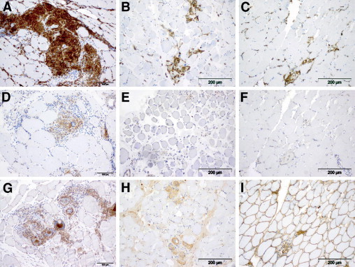
The etiopathogenesis of sarcoidosis, a systemic granulomatous disease, still remains obscure. A multitude of organs have been described to be affected in systemic sarcoidosis. Skeletal muscles may also be affected, leading to myalgia and weakness. A workup of the specific immune response with emphasis on the macrophage response is provided herein. Affected muscle tissue from seven patients with systemic sarcoidosis was analyzed and compared with that from seven patients with other myopathies containing macrophagocytic infiltration. Monocytes/macrophages and giant cells in granulomas of muscle tissue from patients with sarcoidosis show a status of alternative activation (M2) based on their expression of CD206, CD301, arginase-1, and suppressor of cytokine signaling-1 as a consequence of a functionally type 2 helper T cell (Th2)-biased cytokine profile. Significant fibrosis and up-regulation of CCL18 were associated with the M2 phenotype of macrophages. Conversely, up-regulated Th1 cytokines did not result in significant classical activation of macrophages (M1), with poor inducible nitric oxide synthase and cyclooxygenase-2 production. Giant cell formation was further associated with up-regulated expression of DNAX-activating protein of 12 kDa (DAP12; gene symbol TYROBP). Functionally, alternative activation of macrophages on the basis of a Th2-biased immune response may induce clinical symptoms and chronic evolution of neuromuscular sarcoidosis. This is the first characterization of Th2-mediated immune mechanisms in neuromuscular sarcoidosis suggesting that a specific macrophage activation status leading to myofibrosis may be a key event in the pathogenesis of this disease.


Figures

Figure 1
Figure 1
Macrophages, epithelioid cells, and giant cells in granulomas are illustrated by Gömöri stainings (A, B, and C) and H&E stainings (D, E, and F) and nonspecific esterase preparation (G, H, and I) in a patient with sarcoidosis (A, D, and G) and in two control patients: BMD case 1 (B, E, and H) and a patient with necrotizing myopathy (C, F, and I). Monocytes strongly express CD68 (J, K, and L), whereas CD11b is less intensely expressed in the control group (M vs N and O); MHC class I molecules (P) are expressed on monocytes and additionally on the sarcolemma of muscle fibers of patients with muscle sarcoidosis and of those with necrotizing myopathy (P and R), whereas in the BMD case, MHC class I molecules are expressed exclusively on monocytes (Q). Original magnification, ×200.
Figure 2
Figure 2
Macrophages/monocytes, giant cells, and epithelioid cells strongly express the mannose receptor (CD206) (A), and they are also SOCS-1 positive (D). Focal up-regulation of CD206 was found on macrophages from the control group as illustrated: BMD case 1 (B) and necrotizing myopathy (C). Macrophages from the control group, however, did not express SOCS-1 [BMD case 1 (E) and necrotizing myopathy (F)]. Immunohistochemical iNOS staining did not differ significantly in both groups as exemplified by iNOS staining of a muscle sarcoidosis specimen (G) compared with a specimen from the patient with BMD (H) and from a patient with necrotizing myopathy (I). Original magnification, ×200.
Figure 3
Figure 3
Log10 Relative Quantification (RQ) values of mRNA calibrated against expression of the respective molecules in normal muscle specimens. A: Both of the experimental groups show expression of the common IL-4R, IL-4 is expressed significantly higher in the muscle sarcoidosis group, and IL-13 is exclusively expressed in that group. B: Markers of alternative activation are expressed at a significantly higher level. C: Furthermore, expression of DAP12 is also higher in the muscle sarcoidosis group. *P < 0.05, **P < 0.01, ***P < 0.001.
Figure 4
Figure 4
Log10 values of mRNA calibrated against expression of the respective molecules in normal muscle specimens. Expression of IFN-γ and IL-18 was significantly higher in the muscle sarcoidosis group than in the control group (A), whereas the expression of iNOS and cyclooxygenase-2 (COX2), as markers of classical macrophage activation, is not up-regulated in either groups (B). C: Expression of IL-10 and IL-12 was significantly higher in the muscle sarcoidosis group than in the control group. NS, not significant. **P < 0.01, ***P < 0.001.
Figure 5
Figure 5
The chemokine CCL18 is expressed on the sarcolemma of muscle fibers but also in the granulomas (A); also, BMD and necrotizing myopathy muscle specimens showed immunoreactivity on their muscle fibers (B and C). Endomysial and perimysial fibrosis is illustrated by means of elastica van Gieson staining in the sarcoidosis muscle (D). As expected, the BMD muscle (E), but not the necrotizing myopathy muscle (F), shows important fibrosis. Neovascularization is illustrated by the presence of CD31- (G) and smooth muscle actin– (J) expressing vessels, whereas no neovascularization is detectable in the control specimens (H, I, K, and L). Original magnification, × 200. M: Up-regulation of CCL18 can also be demonstrated at the mRNA level (log10 values of mRNA calibrated against the expression of CCL18 in normal muscle specimens are illustrated). ***P < 0.001.

References

    1. Griffith D.E., Aksamit T., Brown-Elliott B.A., Catanzaro A., Daley C., Gordin F., Holland S.M., Horsburgh R., Huitt G., Iademarco M.F., Iseman M., Olivier K., Ruoss S., von Reyn C.F., Wallace R.J., Jr, Winthrop K. An official ATS/IDSA statement: diagnosis, treatment, and prevention of nontuberculous mycobacterial diseases. Am J Respir Crit Care Med. 2007;175:367–416. - PubMed
    1. Grunewald J., Eklund A. Role of CD4+ T cells in sarcoidosis. Proc Am Thorac Soc. 2007;4:461–464. - PMC - PubMed
    1. Morey S.S. American Thoracic Society issues consensus statement on sarcoidosis. Am Fam Physician. 2000;61:553–554. 556. - PubMed
    1. Parrish S., Turner J.F. Diagnosis of sarcoidosis. Dis Mon. 2009;55:693–703. - PubMed
    1. Lazarus A. Sarcoidosis: epidemiology, etiology, pathogenesis, and genetics. Dis Mon. 2009;55:649–660. - PubMed

MeSH terms