. 2011 Mar 2;13(3):274-82.
doi: 10.1016/j.cmet.2011.01.017.

Phosphate incorporation during glycogen synthesis and Lafora disease

Affiliations

Phosphate incorporation during glycogen synthesis and Lafora disease

Vincent S Tagliabracci et al. Cell Metab. .

Abstract

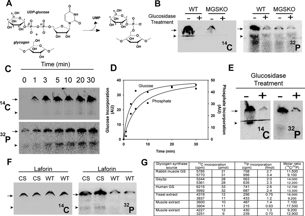
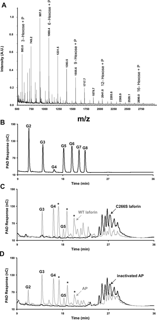
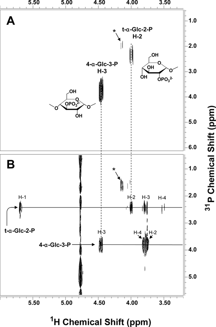
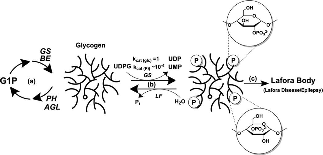
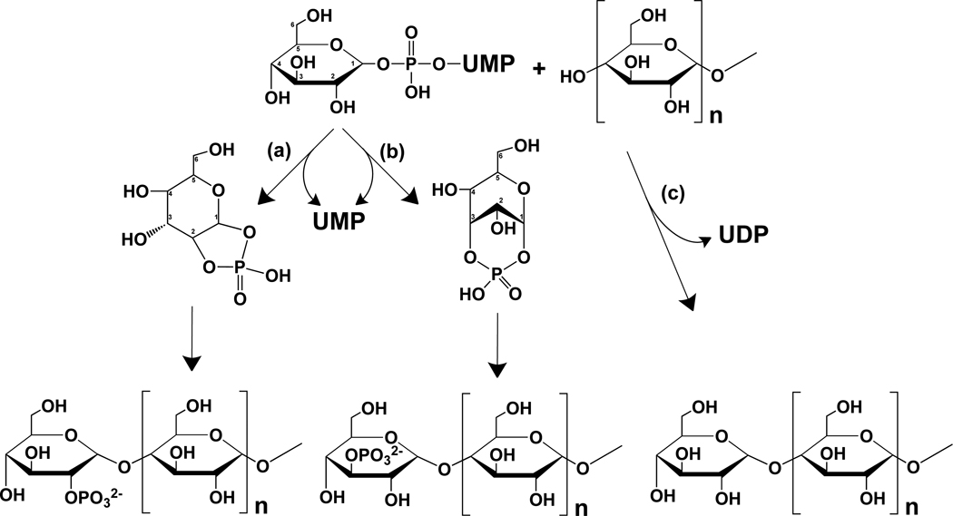
Glycogen is a branched polymer of glucose that serves as an energy store. Phosphate, a trace constituent of glycogen, has profound effects on glycogen structure, and phosphate hyperaccumulation is linked to Lafora disease, a fatal progressive myoclonus epilepsy that can be caused by mutations of laforin, a glycogen phosphatase. However, little is known about the metabolism of glycogen phosphate. We demonstrate here that the biosynthetic enzyme glycogen synthase, which normally adds glucose residues to glycogen, is capable of incorporating the β-phosphate of its substrate UDP-glucose at a rate of one phosphate per approximately 10,000 glucoses, in what may be considered a catalytic error. We show that the phosphate in glycogen is present as C2 and C3 phosphomonoesters. Since hyperphosphorylation of glycogen causes Lafora disease, phosphate removal by laforin may thus be considered a repair or damage control mechanism.


Figures

Figure 1
Figure 1
Glycogen synthase incorporates phosphate into glycogen from UDP-glucose. (A) UDP-glucose structure and the glucose-1-phosphate transferase reaction proposed by Whelan (see text) (B) Incorporation of [U-14C]glucose (left) and 32P (right) into glycogen (6.67 mg/mL) catalyzed by muscle extracts from wild type (WT) mice or mice lacking muscle glycogen synthase (MGSKO) using UDP-[U-14C]glucose and [β-32P]UDP-glucose, respectively, as glucose donors. Reaction products were separated by SDS-PAGE and radioactivity detected by phosphorimaging. The arrow indicates the bottom of the well where glycogen accumulates and the arrowhead the top of the stacking gel where smaller polysaccharides migrate. Some samples were also treated with α-amylase and amyloglucosidase, as indicated, prior to SDS-PAGE. Loss of radioactivity indicates association with polysaccharide. (C) Time dependent incorporation of [U-14C]glucose (upper) and 32P (lower) into glycogen (6.67 mg/mL) by purified rabbit muscle glycogen synthase (1 µg/mL). (D) Quantitation of the phosphorimager data in C. Different gels were analyzed for 32P and 14C incorporation and the arbitrary units are not comparable. Incorporation of radioactivity was quantitated in absolute terms by excision of the relevant gel areas and scintillation counting. (E) Labeled glycogen, prepared as in C, was purified by ethanol precipitation, subjected to treatment with α-amylase and amyloglucosidase, as indicated, and analyzed as in B. (F) Glycogen, prepared as in C, was purified by ethanol precipitation and subjected to treatment with either catalytically inactive C266S laforin (CS) or active laforin (WT). (G) Quantitation of 14C and 32P incorporation into glycogen. Glycogen was synthesized with the indicated source of glycogen synthase and duplicate measurements quantitated as described under Experimental Procedures. See also Figure S1.
Figure 2
Figure 2
Purification and analysis of phosphorylated oligosaccharides from rabbit muscle glycogen. (A) MALDI-TOF mass spectrometry of phosphorylated oligosaccharides (fraction V; see Fig. S1A) from rabbit muscle glycogen. The spectrum displays a series of species differing by 162.1 m/z indicative of a mixture of phosphorylated oligosaccharides ranging from 3 hexoses + phosphate to 16 hexoses + phosphate. (B) HPAEC analysis of malto-oligosaccharide standards. G1, glucose, G2, maltose, G3, maltotriose, G4, maltotetraose, G5, maltopentaose, G6, maltohexaose, G7, maltoheptaose, G8, maltooctaose. (C) Phosphorylated oligosaccharides (fraction V) were treated with either catalytically inactive laforin (C266S, black) of active laforin (WT, grey) and analyzed by HPAEC. (D) Phosphorylated oligosaccharides were treated with either heat denatured alkaline phosphatase (inactivated AP, black) or active alkaline phosphatase (AP, grey) and analyzed by HPAEC. The asterisks mark species generated by laforin or alkaline phosphatase treatment that do not align with any of the linear oligosaccharide standards (panel B) and which are likely branched oligosaccharides. See also Figure S2.
Figure 3
Figure 3
1H-31P correlated NMR spectra of phosphorylated oligosaccharides. The spectra were acquired at 25 °C and 11.7 T with a 1-s relaxation delay and 3.5 kHz spectral width in F2 (A) The 1H-31P gHMQC spectrum was acquired with an 8.0 kHz spectral width in F1, 0.4 s acquisition time, 32 increments, and 360 transients per increment. (B) The 1H-31P HMQC-TOCSY spectrum was acquired with a 3.0 kHz spectral width in F1, 0.3 s acquisition time, 40 ms spinlock time, 24 increments, and 400 transients per increment. Phosphorus decoupling was turned on during acquisition using the GARP-1 decoupling sequence. The signal marked with an asterisk is probably from a terminal glucose-2-phosphate residue linked in a different position but could not be clearly identified due to its low abundance. See also Figure S3.
Figure 4
Figure 4
Model for glycogen phosphate metabolism and Lafora Body formation. (a) Glycogen is synthesized by glycogen synthase (GS) and branching enzyme (BE), and degraded by glycogen phosphorylase (PH) and debranching enzyme (AGL). (b) Glycogen synthase infrequently (1 in ~10,000) incorporates phosphate residues into glycogen. The kcat values given simply denote the relative rates of glucose versus glucose phosphate incorporation. Excessive incorporation of phosphate as C2 or C3 phosphomonesters, which disrupts glycogen structure, is normally kept in check by the action of the laforin phosphatase (LF). (c) When laforin is defective, excessive phosphorylation results in the formation of Lafora bodies and Lafora disease.
Figure 5
Figure 5
Proposed mechanism for the introduction of phosphate into glycogen by glycogen synthase. Glycogen synthase normally transfers a glucose residue from UDP-glucose to form a new α-1,4-glycosidic linkage with the C4-hydroxyl at the reducing end of a glycogen chain (pathway c). The proposed phosphorylation mechanism would involve the formation, in the active site of glycogen synthase, of either glucose-1,2-cyclic phosphate (pathway a) or glucose-1,3-cyclic phosphate (pathway b). Reaction of the C1 of the cyclic phosphodiester would result in the transfer of either a glucose-2-phosphate or a glucose-3-phosphate moiety in α-1,4-glycosidic linkage to a reducing end of glycogen.

Comment in

Similar articles

Cited by

References

    1. Andrade DM, Turnbull J, Minassian BA. Lafora disease, seizures and sugars. Acta Myol. 2007;26:83–86. - PMC - PubMed
    1. Ball S, Guan HP, James M, Myers A, Keeling P, Mouille G, Buleon A, Colonna P, Preiss J. From glycogen to amylopectin: a model for the biogenesis of the plant starch granule. Cell. 1996;86:349–352. - PubMed
    1. Baskaran S, Roach PJ, DePaoli-Roach AA, Hurley TD. Structural basis for glucose-6-phosphate activation of glycogen synthase. Proc Natl Acad Sci U S A. 2010;107:17563–17568. - PMC - PubMed
    1. Blennow A, Nielsen TH, Baunsgaard L, Mikkelsen R, Engelsen SB. Starch phosphorylation: a new front line in starch research. Trends Plant Sci. 2002;7:445–450. - PubMed
    1. Cheng C, Mu J, Farkas I, Huang D, Goebl MG, Roach PJ. Requirement of the self-glucosylating initiator proteins Glg1p and Glg2p for glycogen accumulation in Saccharomyces cerevisiae. Mol Cell Biol. 1995;15:6632–6640. - PMC - PubMed

Publication types

MeSH terms