

The significance of vascular and neural apoptosis to the pathology of diabetic retinopathy
- PMID: 21357409
- PMCID: PMC3053099
- DOI: 10.1167/iovs.10-6293
The significance of vascular and neural apoptosis to the pathology of diabetic retinopathy
Abstract
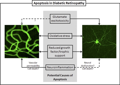
The most striking features of diabetic retinopathy are the vascular abnormalities that are apparent by fundus examination. There is also strong evidence that diabetes causes apoptosis of neural and vascular cells in the retina. Thus, there is good reason to define diabetic retinopathy as a form of chronic neurovascular degeneration. In keeping with the gradual onset of retinopathy in humans, the rate of cell loss in the animal models is insidious, even in uncontrolled diabetes. This is not surprising given that a sustained high rate of cell loss without regeneration would soon lead to catastrophic tissue destruction. The consequences of ongoing cell death are difficult to detect, and even the quantification of cumulative cell loss requires painstaking histology and microscopy. This subtle cell loss raises the issue of the relevance of the phenomenon to the progression of diabetic retinopathy and the ultimate loss of vision. Neuronal function may be compromised in advance of apoptosis, contributing to an early deterioration of vision. Here we review some of the evidence supporting apoptotic cell death as a contributing mechanism of diabetic retinopathy, explore some of the potential causes, and discuss the potential links between apoptosis and loss of visual function in diabetic retinopathy.
Figures
References
-
- Engerman RL, Kern TS. Retinopathy in animal models of diabetes. Diabet Metabol Rev. 1995;11:109–120 - PubMed
-
- Bresnick GH, Davis MD, Myers FL, de Venecia G. Clinicopathologic correlations in diabetic retinopathy, II: clinical and histologic appearances of retinal capillary microaneurysms. Arch Ophthalmol. 1977;95:1215–1220 - PubMed
-
- Sugiyama T, Kobayashi M, Kawamura H, Li Q, Puro DG. Enhancement of P2X(7)-induced pore formation and apoptosis: an early effect of diabetes on the retinal microvasculature. Invest Ophthalmol Vis Sci. 2004;45:1026–1032 - PubMed
-
- Kern TS, Tang J, Mizutani M, et al. Response of capillary cell death to aminoguanidine predicts the development of retinopathy: comparison of diabetes and galactosemia. Invest Ophthalmol Vis Sci. 2000;41:3972–3978 - PubMed
Publication types
MeSH terms
Grants and funding
LinkOut - more resources
Full Text Sources
Other Literature Sources
Medical
