

The cancer/testis antigen prostate-associated gene 4 (PAGE4) is a highly intrinsically disordered protein
- PMID: 21357425
- PMCID: PMC3077599
- DOI: 10.1074/jbc.M110.210765
The cancer/testis antigen prostate-associated gene 4 (PAGE4) is a highly intrinsically disordered protein
Abstract
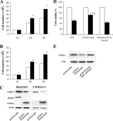
The cancer/testis antigens (CTAs) are an important group of heterogeneous proteins that are predominantly expressed in the testis in the normal human adult but are aberrantly expressed in several types of cancers. Prostate-associated gene 4 (PAGE4) is a member of the CT-X family of CTAs that in addition to testis, is highly expressed in the fetal prostate, and may also play an important role both in benign and malignant prostate diseases. However, the function of this gene remains poorly understood. Here, we show that PAGE4 is a highly (100%) intrinsically disordered protein (IDP). The primary protein sequence conforms to the features of a typical IDP sequence and the secondary structure prediction algorithm metaPrDOS strongly supported this prediction. Furthermore, SDS-gel electrophoresis and analytical size exclusion chromatography of the recombinant protein revealed an anomalous behavior characteristic of IDPs. UV circular dichroism (CD) and NMR spectroscopy confirmed that PAGE4 is indeed a highly disordered protein. In further bioinformatic analysis, the PredictNLS algorithm uncovered a potential nuclear localization signal, whereas the algorithm DBS-Pred returned a 99.1% probability that PAGE4 is a DNA-binding protein. Consistent with this prediction, biochemical experiments showed that PAGE4 preferentially binds a GC-rich sequence. Silencing PAGE4 expression induced cell death via apoptosis and in mice carrying PCa xenografts, siRNA-mediated knockdown of the PAGE4 mRNA attenuated tumor growth in vivo. Furthermore, overexpressing PAGE4 protected cells from stress-induced death. To our knowledge, PAGE4 is the first example of a CTA that is an IDP with an anti-apoptotic function.
Figures


Comment in
-
Re: The cancer/testis antigen prostate-associated gene 4 (PAGE4) is a highly intrinsically disordered protein.J Urol. 2011 Dec;186(6):2494-5. doi: 10.1016/j.juro.2011.08.120. Epub 2011 Oct 26. J Urol. 2011. PMID: 22078626 No abstract available.
Similar articles
-
The Stress-response protein prostate-associated gene 4, interacts with c-Jun and potentiates its transactivation.Biochim Biophys Acta. 2014 Feb;1842(2):154-63. doi: 10.1016/j.bbadis.2013.11.014. Epub 2013 Nov 18. Biochim Biophys Acta. 2014. PMID: 24263171 Free PMC article.
-
Phosphorylation-induced Conformational Ensemble Switching in an Intrinsically Disordered Cancer/Testis Antigen.J Biol Chem. 2015 Oct 9;290(41):25090-102. doi: 10.1074/jbc.M115.658583. Epub 2015 Aug 4. J Biol Chem. 2015. PMID: 26242913 Free PMC article.
-
Cancer/Testis Antigens: "Smart" Biomarkers for Diagnosis and Prognosis of Prostate and Other Cancers.Int J Mol Sci. 2017 Mar 31;18(4):740. doi: 10.3390/ijms18040740. Int J Mol Sci. 2017. PMID: 28362316 Free PMC article. Review.
-
Prostate-associated gene 4 (PAGE4), an intrinsically disordered cancer/testis antigen, is a novel therapeutic target for prostate cancer.Asian J Androl. 2016 Sep-Oct;18(5):695-703. doi: 10.4103/1008-682X.181818. Asian J Androl. 2016. PMID: 27270343 Free PMC article. Review.
-
Cancer/testis antigen PAGE4, a regulator of c-Jun transactivation, is phosphorylated by homeodomain-interacting protein kinase 1, a component of the stress-response pathway.Biochemistry. 2014 Mar 18;53(10):1670-9. doi: 10.1021/bi500013w. Epub 2014 Mar 5. Biochemistry. 2014. PMID: 24559171 Free PMC article.
Cited by
-
Ectopic expression of cancer-testis antigens in cutaneous T-cell lymphoma patients.Clin Cancer Res. 2014 Jul 15;20(14):3799-808. doi: 10.1158/1078-0432.CCR-14-0307. Epub 2014 May 21. Clin Cancer Res. 2014. PMID: 24850846 Free PMC article.
-
GAGE cancer-germline antigens are recruited to the nuclear envelope by germ cell-less (GCL).PLoS One. 2012;7(9):e45819. doi: 10.1371/journal.pone.0045819. Epub 2012 Sep 20. PLoS One. 2012. PMID: 23029259 Free PMC article.
-
Illuminating Intrinsically Disordered Proteins with Integrative Structural Biology.Biomolecules. 2023 Jan 7;13(1):124. doi: 10.3390/biom13010124. Biomolecules. 2023. PMID: 36671509 Free PMC article. Review.
-
PAGE4 and Conformational Switching: Insights from Molecular Dynamics Simulations and Implications for Prostate Cancer.J Mol Biol. 2018 Aug 3;430(16):2422-2438. doi: 10.1016/j.jmb.2018.05.011. Epub 2018 Jun 5. J Mol Biol. 2018. PMID: 29758263 Free PMC article.
-
Prostate-associated gene 4 (PAGE4) protects cells against stress by elevating p21 and suppressing reactive oxygen species production.Am J Clin Exp Urol. 2013 Dec 25;1(1):39-52. eCollection 2013. Am J Clin Exp Urol. 2013. PMID: 25374899 Free PMC article.
References
-
- Hazy E., Tompa P. (2009) ChemPhysChem 10, 1415–1419 - PubMed
-
- Tompa P., Szász C., Buday L. (2005) Trends Biochem. Sci. 30, 484–489 - PubMed
-
- Uversky V. N., Oldfield C. J., Dunker A. K. (2008) Annu. Rev. Biophys. 37, 215–246 - PubMed
-
- Scanlan M. J., Simpson A. J., Old L. J. (2004) Cancer Immun. 4, 1. - PubMed
Publication types
MeSH terms
Substances
Grants and funding
LinkOut - more resources
Full Text Sources
Molecular Biology Databases
Miscellaneous
