. 2011 Mar;60(3):867-75.
doi: 10.2337/db10-1157.

Interleukin-21 is critically required in autoimmune and allogeneic responses to islet tissue in murine models

Affiliations

Interleukin-21 is critically required in autoimmune and allogeneic responses to islet tissue in murine models

Helen M McGuire et al. Diabetes. 2011 Mar.

Abstract

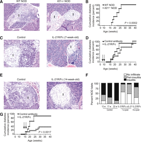
Objective: Type 1 diabetes is an incurable chronic autoimmune disease. Although transplantation of pancreatic islets may serve as a surrogate source of insulin, recipients are subjected to a life of immunosuppression. Interleukin (IL)-21 is necessary for type 1 diabetes in NOD mice. We examined the efficacy of an IL-21-targeted therapy on prevention of diabetes in NOD mice, in combination with syngeneic islet transplantation. In addition, we assessed the role of IL-21 responsiveness in islet allograft rejection in mouse animal models.

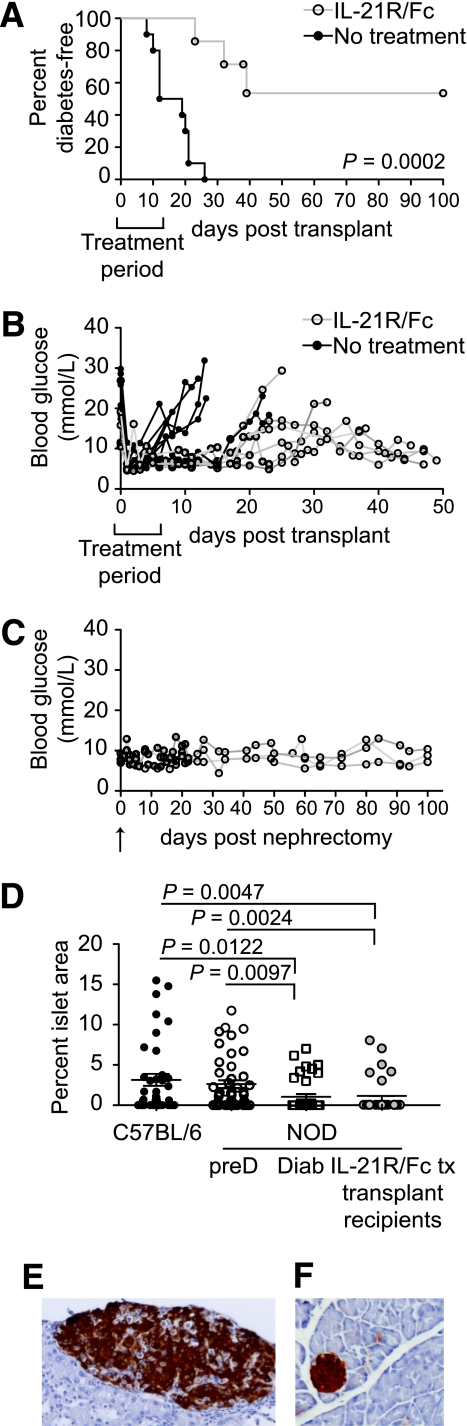
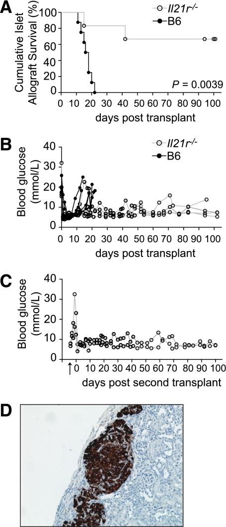
Research design and methods: NOD mice were treated with IL-21R/Fc, an IL-21-neutralizing chimeric protein. This procedure was combined with syngeneic islet transplantation to treat diabetic NOD mice. Survival of allogeneic islet grafts in IL-21R-deficient mice was also assessed.

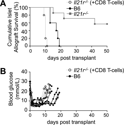
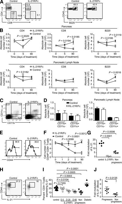
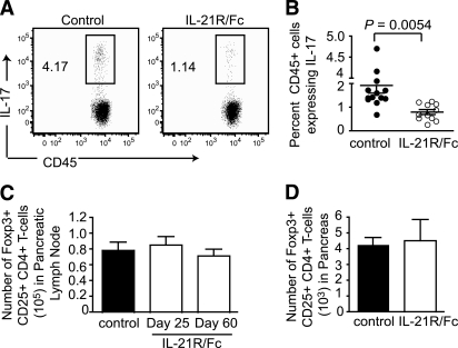
Results: Evidence is provided that IL-21 is continually required by the autoimmune infiltrate, such that insulitis was reduced and reversed and diabetes inhibited by neutralization of IL-21 at a late preclinical stage. Recovery from autoimmune diabetes was achieved by combining neutralization of IL-21 with islet transplantation. Furthermore, IL-21-responsiveness by CD8+ T-cells was sufficient to mediate islet allograft rejection.

Conclusions: Neutralization of IL-21 in NOD mice can inhibit diabetes, and when paired with islet transplantation, this therapeutic approach restored normoglycemia. The influence of IL-21 on a graft-mounted immune response was robust, since the absence of IL-21 signaling prevented islet allograft rejection. These findings suggest that therapeutic manipulation of IL-21 may serve as a suitable treatment for patients with type 1 diabetes.


Figures

FIG. 1.
FIG. 1.
IL-21 helps to maintain pancreatic immune cell infiltrate. A: Sections from paraffin-embedded pancreata were stained with hematoxylin and eosin to reveal mononuclear infiltrate in and around islets of 15-week-old WT NOD and Il21−/− NOD mice, with cumulative incidence of diabetes in WT NOD and Il21−/− NOD mice (B), where n = 12 mice per group. C: Sections from paraffin-embedded pancreata from NOD mice treated at 7 weeks of age with a total of 20 μg control antibody or IL-21R/Fc, given every other day for 11 days are shown at 5 weeks after completion of treatment, with cumulative incidence of diabetes (D), where n = 7 mice per group. Arrows indicate beginning and end of treatment period. E: Sections from paraffin-embedded pancreata from NOD mice treated at 14 weeks of age with a total of 20 μg control antibody or IL-21R/Fc, given every other day for 11 days are shown at 1 week after completion of treatment, with insulitis indices from sections of pancreata showing the percent of islets exhibiting peri-insulitis or insulitis in 12-week-old, 17-week-old, and 22-week-old control mice, and 17-week-old mice treated with IL-21R/Fc or IL-21 polyclonal antibody (1 week after completion of treatment) and 22-week-old mice treated with IL-21R/Fc (6 weeks after completion of treatment) (F), n ≥ 220 islets from 10 mice per group. G: Cumulative incidence of diabetes in NOD mice commencing treatment at 14 weeks of age, where n = 13 mice per group. Arrows indicate beginning and end of treatment period. w, week; WT, wild type. (A high-quality digital representation of this figure is available in the online issue.)
FIG. 4.
FIG. 4.
IL-21 neutralization prolongs survival of autoimmune diabetic mice. A: Percent diabetes-free NOD mice after syngeneic islet transplants with IL-21R/Fc, with treatment period indicated on the graph. n = 10 for control NOD group, no treatment, mean survival time 16.1 days. n = 7 for treated NOD group, three of seven mice returned to a diabetic state. P = 0.0002 (log-rank test). B: Individual blood glucose readings are shown for a cohort of untreated and IL-21R/Fc–treated mice. Treatment period is indicated on the graphs. C: Individual blood glucose readings are shown for long-term surviving IL-21R/Fc–treated NOD mice (n = 3) after nephrectomy (indicated by arrow). D: Frequency of islet mass area in pancreas of long-term IL-21R/Fc–treated NOD syngeneic graft recipients compared with 10-week-old C57BL/6 and prediabetic (preD) NOD mice, and newly diabetic (Diab) NOD mice (15–17 weeks of age). Enumerated from histological sections, with at least 50 fields scored, from three mice per group. Representative histological analyses of a long-term surviving islet graft (E) and pancreas (F) from a long-term surviving IL-21R/Fc–treated NOD mouse. (A high-quality digital representation of this figure is available in the online issue.)
FIG. 5.
FIG. 5.
Islet allografts exhibit prolonged survival in Il21r−/− mice. A: Percent islet graft survival in B6 and Il21r−/− mice following allogenic islet transplants (BALB/c donors). n = 8 for control group, mean survival time 16.6 days. n = 6 for Il21r−/− group. P = 0.0039 (log-rank test). B: Individual blood glucose readings are shown for a cohort of B6 and Il21r−/− mice. C: As indicated by the arrow, nephrectomies were performed on Il21r−/− mice, which survived long-term after receiving an islet allograft. After the removal of the engrafted kidney, blood glucose sharply rose. On day 0 a subsequent islet allograft was transplanted on the contralateral kidney. D: Representative histological analyses of a long-term surviving islet allograft from an Il21r−/− mouse, with insulin staining in brown. (A high-quality digital representation of this figure is available in the online issue.)
FIG. 6.
FIG. 6.
Restoring CD8 T-cell responsiveness to IL-21 results in rapid islet allograft rejection. A: Percent islet graft survival in B6 and Il21r−/− mice, and Il21r−/− mice reconstituted with 1 million IL-21R competent CD8+ T-cells on day −4 (+CD8 T-cells), after allogenic islet transplants (day 0) (BALB/c donors). n = 5 for control B6 group, mean survival time of 16.7 days. n = 6 for Il21r−/− group. n = 5 for Il21r−/− (+CD8 T-cells) group. B: Individual blood glucose readings are shown for a cohort of B6 and Il21r−/− (+CD8 T-cells) mice.
FIG. 2.
FIG. 2.
IL-21 neutralization reduces lymphocyte populations. NOD mice were treated with IL-21R/Fc or control antibody 2.8 μg/i.v. every other day, with a complete dosing of 20 μg achieved on day 11. Cellular composition of PBS-perfused pancreas preparations, and pancreatic lymph nodes were examined by flow cytometric analysis at time points relative to the start of treatment. A: Representative dot plots of CD4+ T-cells, CD8+ T-cells (CD3+ CD45+ gating), and B220+ cells (CD45+ gating) in the pancreas on day 5 (receiving three doses of 2.8 μg). Numbers represent percentage of total lymphocytes. B: Absolute cell number of CD4+ T-cells, CD8+ T-cells, and B220+ cells in the pancreas and pancreatic lymph nodes on day 5 and day 90 (receiving full 20-μg dose) of treatment with IL-21R/Fc. C: Representative dot plots of CD11b and CD11c staining (CD45+ gating) in the pancreas of IL-21R/Fc–treated mice, day 5. D: Absolute cell number of CD11b+ CD11c- macrophages and CD11b+ CD11c+ dendritic cells in NOD pancreas, measured on day 5 of treatment with IL-21R/Fc. Data are presented as means ± SEM, where n = 5–14 for each group, compiled from four experiments. Representative dot plot (E) and absolute cell number of CD44hi CD8+ T-cells from the pancreata of control and IL-21R/Fc–treated NOD mice (F). G: IL-21 mRNA expression measured in pancreas of age-matched control and treated mice on day 3 of treatment with IL-21R/Fc, and NOD mice remaining diabetes free at 40 weeks, termed nonprogressors, where n = 4–5 for each group, from two experiments. IL-21–expressing cells shown as representative dot plots (H) (CD4+ CD3+ CD45+ gating) day 5, and as a percentage of CD4+ T-cells (I), measured in pancreas of control mice and mice on days of treatment with IL-21R/Fc shown, also compared with NOD mice remaining diabetes free at 35 weeks, termed nonprogressors, and diabetic NOD mice, aged 14–22 weeks, where n = 5–10 for each group, from four experiments. J: IL-21 mRNA expression measured by real-time PCR in blood from 12-week-old NOD mice. The cohort were monitored for diabetes onset until 40 weeks of age, and grouped into those that developed disease (progressor) or nonprogressors.
FIG. 3.
FIG. 3.
IL-21 neutralization modulates activated TH subsets. IL-17–expressing cells shown as representative dot plots (A) and quantified in the pancreas of treated vs. control mice on day 3 of treatment with IL-21R/Fc (B), as percentage of CD45+ cells, where n = 12 per group compiled from five experiments. Absolute number of Foxp3+ CD25+ CD4+ T-cells in pancreatic lymph node on days of treatment with IL-21R/Fc shown (C), and in pancreas on day 60 after treatment with IL-21R/Fc (D). Data are presented as means ± SEM, where n = 5 per group from two experiments.

Comment in

References

    1. Anderson MS, Bluestone JA. The NOD mouse: a model of immune dysregulation. Annu Rev Immunol 2005;23:447–485 - PubMed
    1. Bach JF, Mathis D. The NOD mouse. Res Immunol 1997;148:285–286 - PubMed
    1. Green EA, Flavell RA. The initiation of autoimmune diabetes. Curr Opin Immunol 1999;11:663–669 - PubMed
    1. Gazda LS, Charlton B, Lafferty KJ. Diabetes results from a late change in the autoimmune response of NOD mice. J Autoimmun 1997;10:261–270 - PubMed
    1. André-Schmutz I, Hindelang C, Benoist C, Mathis D. Cellular and molecular changes accompanying the progression from insulitis to diabetes. Eur J Immunol 1999;29:245–255 - PubMed

Publication types