Comparative Study
. 2011 Apr 1;186(7):4019-26.
doi: 10.4049/jimmunol.1003566. Epub 2011 Feb 25.

β1 integrin is critical for the maintenance of antigen-specific CD4 T cells in the bone marrow but not long-term immunological memory

Affiliations
Comparative Study

β1 integrin is critical for the maintenance of antigen-specific CD4 T cells in the bone marrow but not long-term immunological memory

Christopher C DeNucci et al. J Immunol. .

Abstract

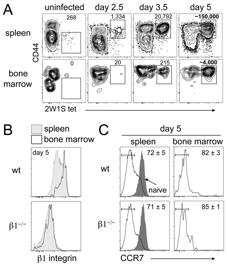
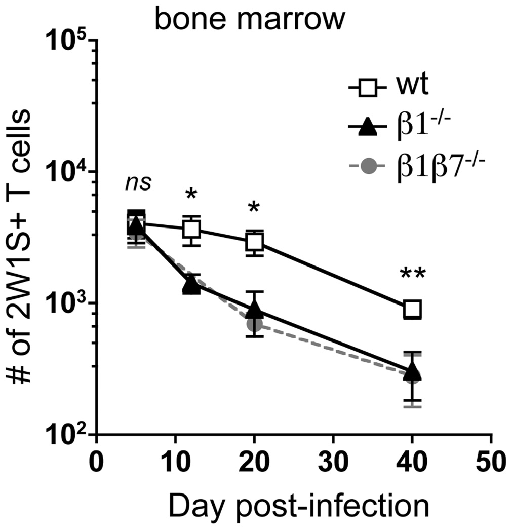
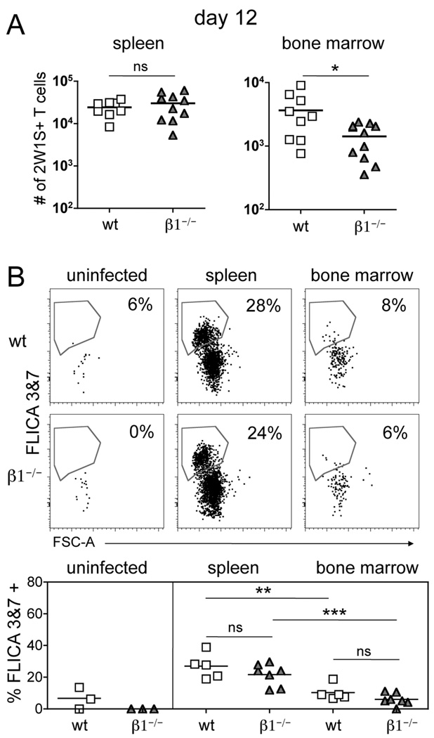
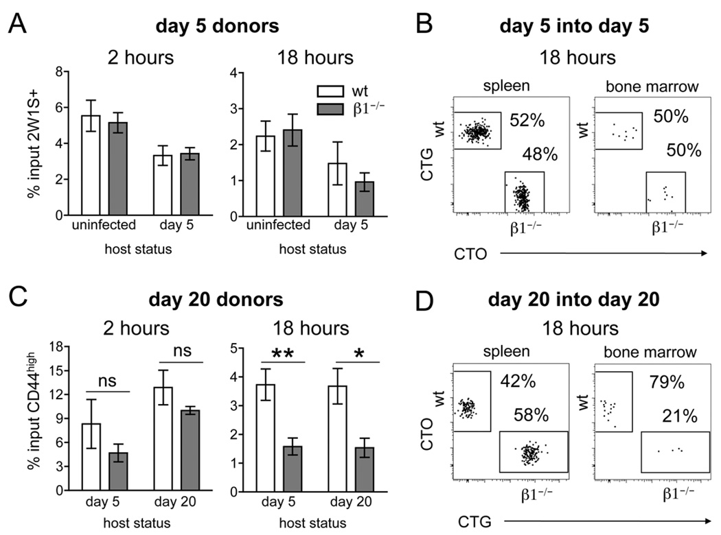
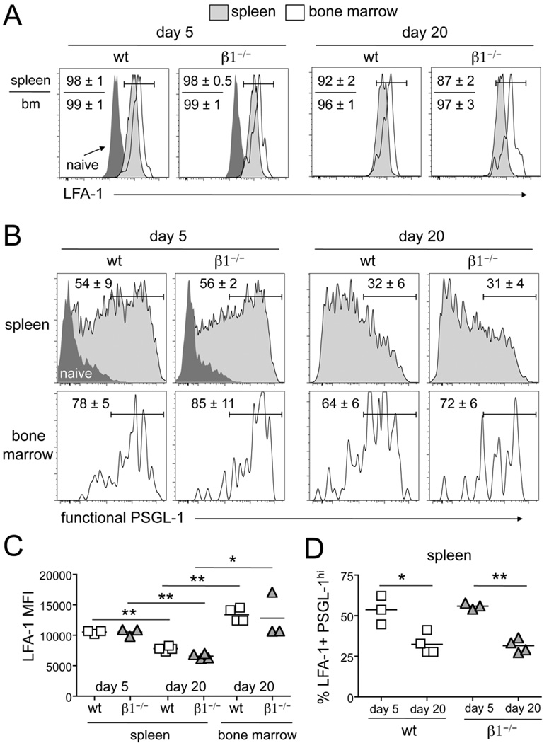
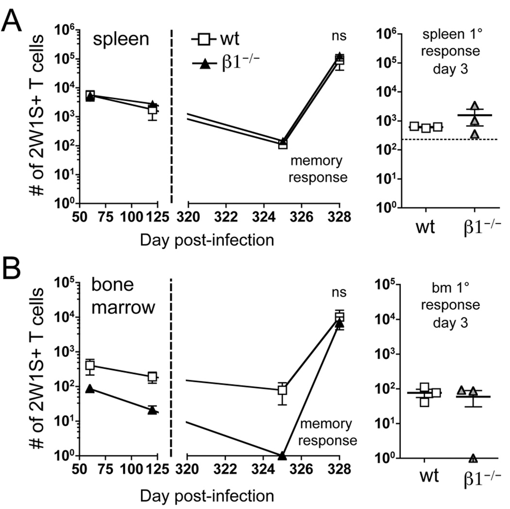
The long-term maintenance of memory CD4 T cells promotes protective immunity against future pathogen reinfection. As a site rich in survival cytokines, the bone marrow is proposed to be a critical niche for the survival of memory CD4 T cells. We demonstrate that endogenous, polyclonal Ag-specific CD4 T cells rapidly enter and are recovered long-term from the bone marrow following i.v. infection with Listeria monocytogenes. β(1) integrin-deficient CD4 T cells also populate the bone marrow early following an infection, but their numbers in this site rapidly decline. This decline was not caused by increased death of T cells lacking β(1) integrin but rather by reduced retention in the bone marrow after the primary immune response. The loss of memory CD4 T cells from the bone marrow does not lead to a loss of the predominant source of memory CD4 T cells in the spleen or the ability to mount a memory response. Thus, β(1) integrin-dependent maintenance of memory CD4 T cells in the bone marrow is not required for long-term CD4 T cell memory.


Figures

Figure 1
Figure 1. CD4 T cells rapidly enter the bone marrow following i.v bacterial infection
(A) Tracking the 2W1S-specific population following i.v. infection with ALm-2W1S in wt mice. Numbers indicate calculated cell number from gate (day 5 are averages, n=9). (B) Spleen and bone marrow histograms of β1 integrin expression on 2W1S-specific CD44high CD4 T cells day 5 post-infection from wt and β1−/− mice. (C) Spleen and bone marrow histograms of CCR7 expression on 2W1S-specific CD44high CD4 T cells day 5 post-infection from wt and β1−/− mice. Gray histogram represents naïve CD44low CD4 T cells from the spleen. Numbers indicate percent of cells in the drawn gate (n=3, mean +/− s.d.).
Figure 2
Figure 2. Maintenance, but not entry, of CD4 T cells in the bone marrow is dependent on β1 integrin
2W1S-specific CD44high CD4 T cells recovered from the bone marrow of wt, β1−/−, and β1β7−/− mice following ALm-2W1S infection. Data are from ten experiments (n=3–11, mean +/– s.e.m.). (ns) p > 0.05, *p < 0.05, **p=0.009 compared to wt, two-tailed unpaired t test.
Figure 3
Figure 3. Loss of CD4 T cells from the bone marrow is not related to increased cell death
(A) Number of 2W1S-specific CD44high CD4 T cells recovered from the spleen and bone marrow 12 days post-infection with ALm-2W1S in wt and β1−/− mice (mean). * p < 0.05, two-tailed unpaired t test. (B) Staining for active caspases 3 & 7 (FLICA 3&7) in 2W1S-specific CD44low CD4 T cells from the spleen of uninfected mice or 2W1S-specific CD44high CD4 T cells from the spleen and bone marrow 12 days post-infection. Bottom graphs show replicates (mean). (ns) p > 0.05, ** p<0.01, *** p<0.001, one-way ANOVA, followed by Tukey’s multiple comparison test.
Figure 4
Figure 4. Loss of CD4 T cells from the bone marrow is due to decreased ability to recirculate
(A) Bar graphs showing the percentage of transferred 2W1S-specific CD44high CD4 T cells recovered from the bone marrow of host mice at 2 and 18 hours post-transfer. Percentage of input was calculated by dividing recovered cell number by transferred cell number (mean +/− s.e.m., n=4–6). (B) Representative dot plots of spleen and bone marrow of mice infected 5 days prior to co-transfer of day 5 post-infection wt and β1−/− 2W1S-specific CD44high CD4 T cells. Transferred cells are distinguished from the host population by labeling with CTG (wt) and CTO (β1−/−). (C) Bar graphs showing the percentage of transferred CD44high CD4 T cells recovered from the bone marrow of host mice at 2 and 18 hours post-transfer (n=4–5, mean +/− s.e.m.). * p=0.02, ** p=0.008, two-tailed unpaired t test. (D) Representative dot plots of spleen and bone marrow of host mice infected 20 days prior to co-transfer of day 20 post-infection wt and β1−/− CD44high CD4 T cells. Transferred cells are distinguished from the host population by labeling with CTO (wt) and CTG (β1−/−). The 2W1S-specific host population (negative for CTO and CTG) has been excluded from all dot plots.
Figure 5
Figure 5. Expression of LFA-1 and functional PSGL-1 is decreased between day 5 and 20 post-infection
2W1S-specific CD44high CD4 T cells from the spleen and bone marrow of wt and β1−/− mice following i.v. infection with ALm-2W1S stained for (A) LFA-1 or (B) functional PSGL-1. Dark gray histograms represent staining of CD44low CD4 T cells (naïve) from the spleen. Numbers indicate percent of cells in the drawn gate (n=3–4, mean +/− s.d.). (C) Median Fluorescence intensity (MFI) of LFA-1 staining on PSGL-1hi 2W1S-specific CD44highCD4 T cells from the spleen and bone marrow of wt and β1−/− mice following i.v. infection with ALm-2W1S. (D) Percentage of LFA-1+ PSGL-1hi 2W1S-specific CD44high CD4 T cells from the spleen and bone marrow of wt and β1−/− mice following i.v. infection with ALm-2W1S. Bar represents the mean. * p<0.02, ** p<0.0002, two-tailed unpaired t test.
Figure 6
Figure 6. Retention of CD4 T cells in the bone marrow is not required for survival or response of memory CD4 T cells
2W1S-specific CD44high CD4 T cells recovered from the spleen (A) and bone marrow (B) of wt and β1−/− mice following i.v. infection with ALm-2W1S. At day 325 post-infection, mice were re-challenged with 2W1S-LPS. Spleen and bone marrow were harvested three days following re-challenge (n=5, mean+/− s.e.m.). The primary (1°) response of naïve (uninfected) mice to 2W1S-LPS was also characterized three days following challenge. Horizontal dotted lines on graphs indicate the average number of 2W1S-specific CD44low CD4 T cells in naïve mice. (ns) p > 0.05, two-tailed unpaired t test.

Similar articles

Cited by

References

    1. Reinhardt RL, Khoruts A, Merica R, Zell T, Jenkins MK. Visualizing the generation of memory CD4 T cells in the whole body. Nature. 2001;410:101–105. - PubMed
    1. Masopust D, Vezys V, Marzo AL, Lefrancois L. Preferential localization of effector memory cells in nonlymphoid tissue. Science. 2001;291:2413–2417. - PubMed
    1. Di Rosa F. T-lymphocyte interaction with stromal, bone and hematopoietic cells in the bone marrow. Immunol. Cell Biol. 2009;87:20–29. - PubMed
    1. Benner R, Meima F, van der Meulen GM. Antibody formation in mouse bone marrow. II. Evidence for a memory-dependent phenomenon. Cell. Immunol. 1974;13:95–106. - PubMed
    1. Price PW, Cerny J. Characterization of CD4+ T cells in mouse bone marrow. I. Increased activated/memory phenotype and altered TCR Vβ repertoire. Eur. J. Immunol. 1999;29:1051–1056. - PubMed

Publication types

MeSH terms