. 2011 Apr 22;286(16):14445-54.
doi: 10.1074/jbc.M110.214635. Epub 2011 Feb 27.

Terminal alkene formation by the thioesterase of curacin A biosynthesis: structure of a decarboxylating thioesterase

Affiliations

Terminal alkene formation by the thioesterase of curacin A biosynthesis: structure of a decarboxylating thioesterase

Jennifer J Gehret et al. J Biol Chem. .

Abstract

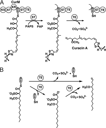
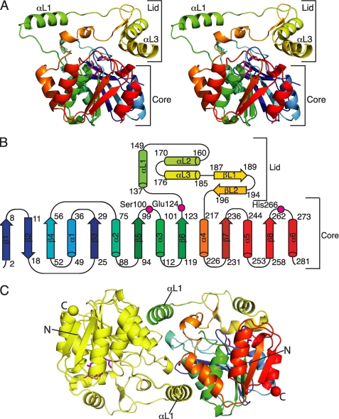
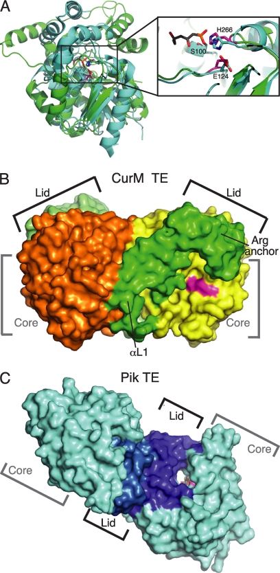
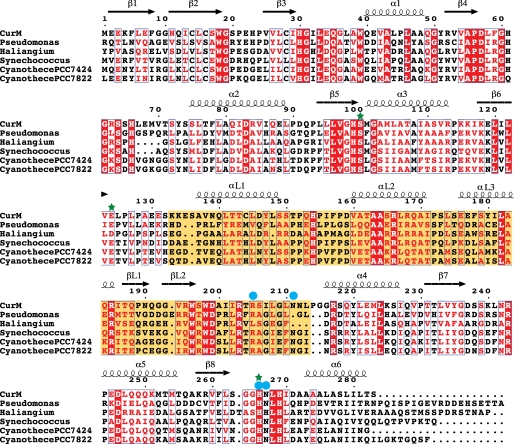
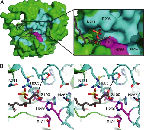
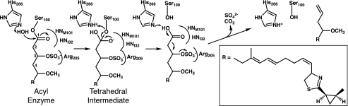
Curacin A is a polyketide synthase (PKS)-non-ribosomal peptide synthetase-derived natural product with potent anticancer properties generated by the marine cyanobacterium Lyngbya majuscula. Type I modular PKS assembly lines typically employ a thioesterase (TE) domain to off-load carboxylic acid or macrolactone products from an adjacent acyl carrier protein (ACP) domain. In a striking departure from this scheme the curacin A PKS employs tandem sulfotransferase and TE domains to form a terminal alkene moiety. Sulfotransferase sulfonation of β-hydroxy-acyl-ACP is followed by TE hydrolysis, decarboxylation, and sulfate elimination (Gu, L., Wang, B., Kulkarni, A., Gehret, J. J., Lloyd, K. R., Gerwick, L., Gerwick, W. H., Wipf, P., Håkansson, K., Smith, J. L., and Sherman, D. H. (2009) J. Am. Chem. Soc. 131, 16033-16035). With low sequence identity to other PKS TEs (<15%), the curacin TE represents a new thioesterase subfamily. The 1.7-Å curacin TE crystal structure reveals how the familiar α/β-hydrolase architecture is adapted to specificity for β-sulfated substrates. A Ser-His-Glu catalytic triad is centered in an open active site cleft between the core domain and a lid subdomain. Unlike TEs from other PKSs, the lid is fixed in an open conformation on one side by dimer contacts of a protruding helix and on the other side by an arginine anchor from the lid into the core. Adjacent to the catalytic triad, another arginine residue is positioned to recognize the substrate β-sulfate group. The essential features of the curacin TE are conserved in sequences of five other putative bacterial ACP-ST-TE tridomains. Formation of a sulfate leaving group as a biosynthetic strategy to facilitate acyl chain decarboxylation is of potential value as a route to hydrocarbon biofuels.


Figures

FIGURE 1.
FIGURE 1.
Chain termination in curacin A biosynthesis. A, off-loading reactions in the final module, CurM. Following extension by the CurM ketosynthase (KS) and acyltransferase (AT) and reduction by the ketoreductase (KR), ST catalyzes transfer of sulfonate to the β-hydroxyl followed by TE hydrolysis of the thioester with concomitant decarboxylation and sulfate elimination. The sulfonate group donor is the PAPS cofactor, which is converted to PAP. B, experimental scheme for the assay of CurM TE. Recombinant CurM ACP loaded with a substrate analog and sulfonated by ST was reacted with excised CurM TE. Activity was monitored by HPLC of the reaction mixture and detection of the holo-ACP product.
FIGURE 2.
FIGURE 2.
Structure of curacin A thioesterase. A, CurM TE polypeptide. The stereo ribbon diagram is colored as a rainbow from blue at the N terminus to red at the C terminus with the catalytic triad residues in stick form with a magenta C. B, topology diagram. CurM TE has an α/β-hydrolase fold in the core domain and a novel lid topology. Residues of the catalytic triad (Ser100, Glu124, and His266) are labeled. C, backbone trace of the CurM TE dimer viewed along the molecular dyad. Monomers are colored as a rainbow (right) and in yellow (left), with the catalytic triad as in A, and N and C termini shown as spheres of the same color as the terminal residue.
FIGURE 3.
FIGURE 3.
Comparison of curacin and pikromycin TEs. A, structure alignment of the core of CurM TE (green) and Pik TE (cyan, Protein Data Bank code 2H7X, (12)) (root mean square deviation = 1.5 Å for 95 Cα atoms). Both structures have the conserved α/β-hydrolase core, but the lids differ. The zoom view shows the active site conservation in the catalytic triad of CurM TE (magenta) and Pik TE (cyan) with a triketide affinity label (gray). The view is similar to Fig. 2A. B, surface representation of the CurM TE dimer. The primary dimer contact is between the lid (subunits in two shades of green) and core (subunits in yellow and orange). The active site (magenta) is in an open cleft between the core and lid. C, surface representation of the Pik TE dimer. The dimer contact is exclusively between the lid subdomains (subunits in two shades of blue) with no contacts of core domains (cyan). The active site (magenta) with an affinity label (gray sticks) is at the center of an open-ended tunnel (12). The views in B and C highlight the differences in active site access, which are distinct for the CurM TE and Pik TE enzymes.
FIGURE 4.
FIGURE 4.
Sequence alignment of CurM TE with putative TEs from open reading frames encoding tandem ACP-ST-TE tridomains. Invariant residues are highlighted in red, conserved residues are printed in red. The conserved lid domain is highlighted in yellow. Green stars indicate active site residues and blue circles indicate residues probed by site-directed mutagenesis. Sequence alignment was performed by MUSCLE (33), and the figure was prepared using ESPript (42). GenBank entries are: Pseudomonas entomophila L48 (GenBank YP_610919), H. ochraceum DSM 14365 (YP_003265308), Synechococcus PCC 7002 (YP_001734428), Cyanothece PCC 7424 (YP_002377174), and Cyanothece PCC 7822 (ZP_03153601).
FIGURE 5.
FIGURE 5.
CurM TE active site. A, surface of the active site cleft. In the molecular surface view, the active site (magenta catalytic triad) is in a cleft in which the presumed phosphopantetheine entrance is lined with conserved residues (cyan), with other parts of the protein surface in green and the modeled acyl-enzyme intermediate in gray sticks. B, modeled acyl-enzyme intermediate (gray C). In this stereo view, the intermediate is surrounded by conserved amino acid residues (cyan C) and the catalytic triad (Ser100, Glu124, and His266) (magenta C). The carbonyl oxygen is bound in the oxyanion hole (hydrogen bonds to the NHs of the Ile32 and Met101).
SCHEME 1.
SCHEME 1.
Predicted reaction sequence for CurM TE hydrolysis, sulfate group elimination, and decarboxylation. Arg205 recognizes the (R)-β-sulfate, facilitating hydrolysis by the catalytic triad. The catalytic His266 is positioned to assist in decarboxylation followed by sulfate elimination.

References

    1. Newman D. J., Cragg G. M. (2007) J. Nat. Prod. 70, 461–477 - PubMed
    1. Fortman J. L., Chhabra S., Mukhopadhyay A., Chou H., Lee T. S., Steen E., Keasling J. D. (2008) Trends Biotechnol. 26, 375–381 - PubMed
    1. Verdier-Pinard P., Lai J. Y., Yoo H. D., Yu J., Marquez B., Nagle D. G., Nambu M., White J. D., Falck J. R., Gerwick W. H., Day B. W., Hamel E. (1998) Mol. Pharmacol. 53, 62–76 - PubMed
    1. Chang Z., Sitachitta N., Rossi J. V., Roberts M. A., Flatt P. M., Jia J., Sherman D. H., Gerwick W. H. (2004) J. Nat. Prod. 67, 1356–1367 - PubMed
    1. Gu L., Geders T. W., Wang B., Gerwick W. H., Håkansson K., Smith J. L., Sherman D. H. (2007) Science 318, 970–974 - PubMed

Publication types

Associated data