Review
. 2011 May;4(3):477-86.
doi: 10.1093/mp/ssr006. Epub 2011 Feb 28.

Transport and metabolism of the endogenous auxin precursor indole-3-butyric acid

Affiliations
Review

Transport and metabolism of the endogenous auxin precursor indole-3-butyric acid

Lucia C Strader et al. Mol Plant. 2011 May.

Abstract

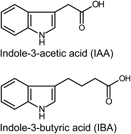
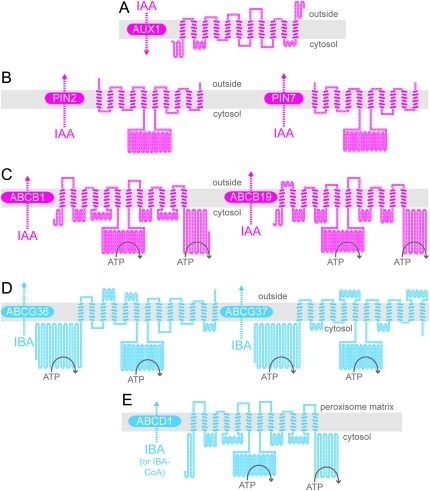
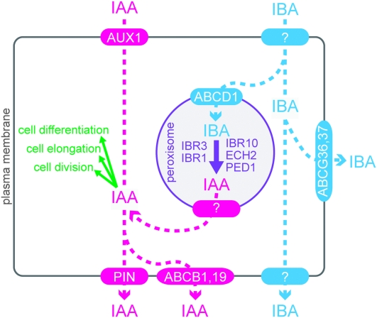
Plant growth and morphogenesis depend on the levels and distribution of the plant hormone auxin. Plants tightly regulate cellular levels of the active auxin indole-3-acetic acid (IAA) through synthesis, inactivation, and transport. Although the transporters that move IAA into and out of cells are well characterized and play important roles in development, little is known about the transport of IAA precursors. In this review, we discuss the accumulating evidence suggesting that the IAA precursor indole-3-butyric acid (IBA) is transported independently of the characterized IAA transport machinery along with the recent identification of specific IBA efflux carriers and enzymes suggested to metabolize IBA. These studies have revealed important roles for IBA in maintaining IAA levels and distribution within the plant to support normal development.


Figures

Figure 1.
Figure 1.
IAA and IBA. Indole-3-acetic acid (IAA) is a naturally occurring auxin. Indole-3-butyric acid (IBA) is a naturally occurring IAA precursor.
Figure 2.
Figure 2.
Predicted Topologies of IAA Carrier Proteins that Do Not Transport IBA (A–C) and Proteins Demonstrated (D) or Suggested (E) to Transport IBA. Schematic diagrams illustrating the predicted topologies of AUX1 (A), PIN2 and PIN7 (B), ABCB1 and ABCB19 (C), ABCG36 and ABCG37 (D), and ABCD1 (E) based on the outputs of ARAMEMNON (http://aramemnon.botanik.uni-koeln.de; Schwacke et al., 2003) and TOPO2 (www.sacs.ucsf.edu/TOPO2). Each amino acid residue is represented by a circle; filled circles represent residues predicted to span the membrane (gray rectangle). Positions of nucleotide-binding domains in the ABC proteins are schematized by ATP hydrolysis. For AUX1, the ARAMEMNON TmConsens prediction of transmembrane domains predicted 10 transmembrane domains with strong scores and one additional transmembrane domain with a weak score. Because AUX1 was experimentally shown to have 11 transmembrane domains (Swarup et al., 2004), all 11 transmembrane domains are depicted in the diagram. We used the ARAMEMNON TmConsens predictions for PIN2, PIN7, ABCB1, ABCB19, and ABCG37 to create the corresponding models, and the ARAMEMNON TmHMM_v2 prediction (Sonnhammer et al., 1998) to create the ABCG36 diagram. The ARAMEMNON MemSat_v3 (Jones et al., 1994) predicted 13 transmembrane domains for ABCD1. However, because the ATPase domains of ABCD1 are cytosolic (Nyathi et al., 2010), we did not include the first predicted transmembrane domain in the ABCD1 diagram.
Figure 3.
Figure 3.
ABCG37 and ABCG36 Localize to the Outer Polar Domain of Root Epidermal and Lateral Root Cap Cells. Confocal images of root tips of 5-day-old seedlings carrying GFP–ABCG37 (pis1-1 carrying 35S:GFP–ABCG37) (Łangowski et al., 2010) and ABCG36–GFP (pen3-1 carrying PEN3:PEN3–GFP) (Stein et al., 2006). The upper panels show GFP signal and the lower panels show propidium iodide staining of endodermal (en), cortex (co), epidermal (ep), and lateral root cap (LRC) cell walls. Both GFP–ABCG37 (Łangowski et al., 2010; Růžička et al., 2010) and ABCG36–GFP (Strader and Bartel, 2009) accumulate on the outward face of epidermal and lateral root cap cells, a polar domain termed the ‘outer polar domain’ (Łangowski et al., 2010), implying that these transporters move IBA from the root into the rhizosphere.
Figure 4.
Figure 4.
Cellular Model Showing Distinct IAA and IBA Transporters. The auxin activity of IBA requires its conversion to IAA in peroxisomes, probably by β-oxidation enzymes encoded by IBR1, IBR3, IBR10, ECH2, and PED1.

References

    1. Bailly A, Sovero V, Vincenzetti V, Santelia D, Bartnik D, Koenig BW, Mancuso S, Martinoia E, Geisler M. Modulation of P-glycoproteins by auxin transport inhibitors is mediated by interaction with immunophilins. J. Biol. Chem. 2008;283:21817–21826. - PubMed
    1. Bandyopadhyay A, et al. Interactions of PIN and PGP auxin transport mechanisms. Biochem. Soc. Trans. 2007;35:137–141. - PubMed
    1. Bednarek P, et al. A glucosinolate metabolism pathway in living plant cells mediates broad-spectrum antifungal defense. Science. 2009;323:101–106. - PubMed
    1. Blakeslee JJ, et al. Interactions among PIN-FORMED and P-glycoprotein auxin transporters in Arabidopsis. Plant Cell. 2007;19:131–147. - PMC - PubMed
    1. Blommaert K. Growth- and inhibiting-substances in relation to the rest period of the potato tuber. Nature. 1954;174:970–972. - PubMed

Publication types