. 2011 May 6;286(18):16272-84.
doi: 10.1074/jbc.M110.195347. Epub 2011 Feb 28.

Precursor of brain-derived neurotrophic factor (proBDNF) forms a complex with Huntingtin-associated protein-1 (HAP1) and sortilin that modulates proBDNF trafficking, degradation, and processing

Affiliations

Precursor of brain-derived neurotrophic factor (proBDNF) forms a complex with Huntingtin-associated protein-1 (HAP1) and sortilin that modulates proBDNF trafficking, degradation, and processing

Miao Yang et al. J Biol Chem. .

Abstract

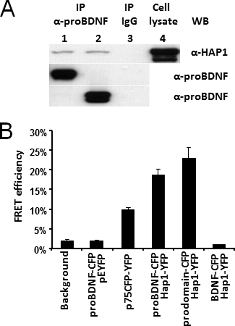
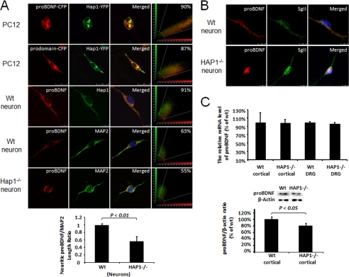
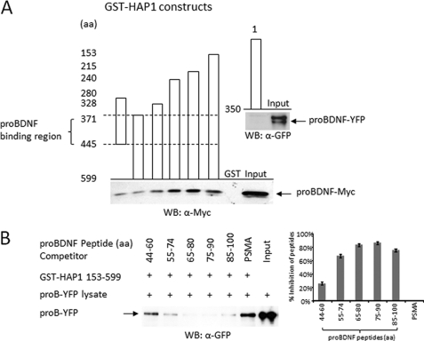
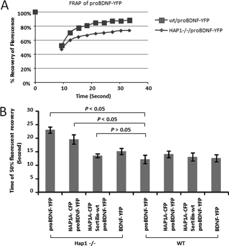
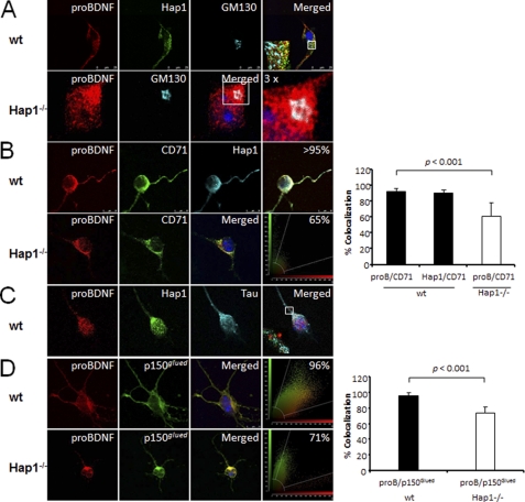
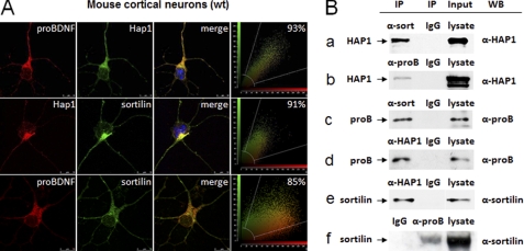
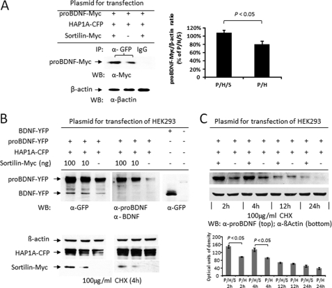
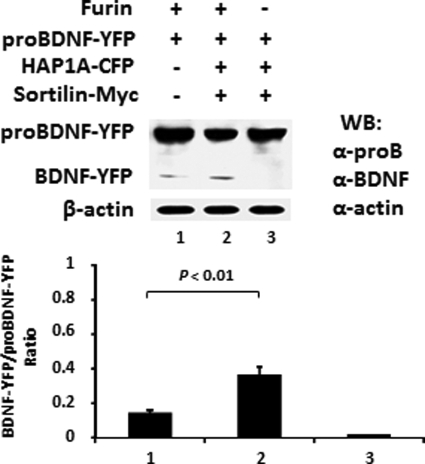
proBDNF, a precursor of brain-derived neurotrophic factor (BDNF), is anterogradely transported and released from nerve terminals, but the mechanism underlying this process remains unclear. In this study, we report that proBDNF forms a complex with Huntingtin associated protein-1 (HAP1) and sortilin, which plays an important role in proBDNF intracellular trafficking and stabilization. The interaction of proBDNF with both HAP1A and sortilin in co-transfected HEK293 cells is confirmed by both fluorescence resonance energy transfer and co-immunoprecipitation. The frequent co-localization (>90%) of endogenous HAP1, sortilin, and proBDNF is also found in cultured cortical neurons. Mapping studies using GST pulldown and competition assays has defined the interacting region of HAP1 with proBDNF within amino acids 371-445 and the binding sequences of proBDNF to HAP1 between amino acids 65 and 90. Fluorescence recovery after photobleaching confirms the defective movement of proBDNF-containing vesicles in neurites of HAP1(-/-) neurons, which can be partially restored by reintroducing HAP1 cDNA into the neurons. However, the effect is significantly increased by simultaneously reintroducing both HAP1 and sortilin. proBDNF and HAP1 are highly co-localized with organelle markers for the Golgi network, microtubules, molecular motor, or endosomes in normal neurons, but this co-localization is reduced in HAP1(-/-) neurons. Co-immunoprecipitation and Western blot showed that sortilin stabilizes the proBDNF·HAP1 complex in co-transfected HEK293 cells, helping to prevent proBDNF degradation. Furthermore, the complex facilitates furin cleavage to release mature BDNF.


Figures

FIGURE 1.
FIGURE 1.
The prodomain of BDNF interacts with HAP1. A, proBDNF and the prodomain interact with HAP1 in co-transfected HEK293 cells. HEK293 co-transfected with HAP1A-YFP·proBDNF-CFP (lane 1) or HAP1A-YFP·prodomain-CFP (lane 2), respectively, were used for immunoprecipitation. Lane 1 and 2, Co-IP with sheep anti-prodomain antibodies (α-proBDNF) and Western blot (WB) with mouse anti-HAP1 (α-HAP1); HAP1A-YFP was detected (top panel). proBDNF-CFP and prodomain-CFP were blotted from the same samples used for Co-IP (middle and bottom panel), respectively. Lane 3, Co-IP with sheep IgG for negative control; no targeted protein was detected from the lysates used in lanes 1 and 2. Lane 4, co-transfected cell lysate input for probing HAP1A-YFP. B, FRET analysis of proBDNF interaction with HAP1A in co-transfected PC12 cells. The proBDNF-CFP·pEYFP were performed for the negative control. The p75CFP-YFP plasmid was used as a positive control. FRETeff was determined from a photobleached and targeted vesicle. The bars represent the means ± S.E. (n = 6).
FIGURE 2.
FIGURE 2.
HAP1 is physiologically associated with proBDNF. A, proBDNF co-localization with HAP1 in co-transfected PC12 cells and mouse cortical neurons. PC12 cells were co-transfected with proBDNF-CFP (red) and HAP1A-YFP (green) (top row). PC12 cells were co-transfected with prodomain of BDNF-CFP, designed as prodomain-CFP (red) and HAP1A-YFP (green) (second row). Mouse WT cortical neurons (third row) were immunostained for proBDNF (red) and HAP1 (green). Mouse WT cortical neurons (fourth row) were immunostained for proBDNF (red) and MAP2 (green). Mouse HAP1 −/− cortical neurons (bottom row) were immunostained for proBDNF (red) and MAP2 (green). HAP1 co-localization with proBDNF or the prodomain was performed using a SP5 confocal microscope and indicated in merged panels (yellow). Snapshots of co-localization are shown in the right panels. The transport of proBDNF in neurites was quantitatively determined by measuring the distance from the soma of proBDNF fluorescence as a ratio of total neurite length (MAP2) (bottom panel). B, HAP1-mediated proBDNF transport is unrelated to secretogranin II vesicle. Mouse WT (upper panel) and HAP1 −/− (bottom panel) cortical neurons immunostained for proBDNF (red) and secretogranin II (SgII, green). C, real time quantitative RT-PCR of proBDNF mRNA level (top). The mRNA expression level was determined in Hap1−/− cortex or dorsal root ganglia (n = 3, p > 0.05). Western blot analysis of proBDNF protein level in cortex of WT and HAP1−/− (bottom). The protein level of proBDNF was significantly lower in HAP1−/− samples (n = 3, p < 0.05).
FIGURE 3.
FIGURE 3.
Mapping proBDNF and HAP1 interacting region. A, GST-HAP1 pulldown assay for mapping proBDNF binding region. The columns indicate seven GST-HAP1 constructs (HAP1 1–350, HAP1 280–445, HAP1 371–599, HAP1 328–599, HAP1 240–599, HAP1 215–599, and HAP1 153–599). proBDNF-YFP lysate was used for incubation with GST-HAP1 1–350 fusion protein and proBDNF-Myc lysate for incubation with all other GST-HAP1 fusion proteins in GST-pulldown assays. proBDNF-YFP used for GST-HAP1 1–350 pulldown assay is indicated by an arrow (top) and blotted with rabbit anti-GFP antibody (a-GFP). proBDNF-Myc used for all other GST-HAP1 pulldown assays is indicated by an arrow from pulled down and input samples (bottom), which was blotted with mouse anti-Myc antibody (a-Myc). The proBDNF binding region is indicated between dashed lines. B, the proBDNF peptides competition assay. Five specific proBDNF peptides (proBDNF 44–60, proBDNF 55–74, proBDNF 65–80, proBDNF 75–90, and proBDNF 85–100) and one nonspecific PSMA peptide used for competing proBDNF-YFP binding to GST-HAP1 153–599 in GST pulldown assay are listed at the top. The positive band of proBDNF-YFP is shown in the Input lane. The arrow indicates proBDNF-YFP either pulled-down by GST-HAP1 153–599 in the competition assay or input samples. On the right, the average inhibition of the proBDNF peptides on the interaction between proBDNF-YFP and GST-HAP1 153–599 from three individual experiments is indicated as a percentage over the nonspecific PSMA peptide, which was calculated as 0% inhibition. WB, Western blot.
FIGURE 4.
FIGURE 4.
Lack of HAP1 impacts on the trafficking of proBDNF-YFP in mouse cortical neurons. A, FRAP of proBDNF-YFP trafficking in living neurons. Mouse cortical neurons were transfected with proBDNF-YFP. The trafficking of proBDNF-YFP in neurites was examined using FRAP under a Leica SP5 confocal microscope. The initial fluorescence is indicated as 100%. B, time for 50% fluorescence recovery was determined by FRAP in transfected mouse cortical neurons. The four bars on the left are in HAP1−/− neurons, and the bars on the right are in WT neurons. The data are represented as the means ± S.E. from six transfected neurons. The labels under each bar indicate different plasmids that are used for transfection. Less time is needed for WT neurons to get a recovery of the fluorescence of proBDNF-YFP compared with HAP1−/− neurons. Co-transfection of HAP1-CFP in HAP1−/− neurons, but not in WT neurons, reduced the time needed for recovery, whereas co-transfection of both HAP1-CFP and sortilin completely rescued the defect in fluorescence recovery.
FIGURE 5.
FIGURE 5.
Differential distribution of organelle markers in HAP1−/− cortical neurons. A, top panel, proBDNF and HAP1 are present in cis-Golgi apparatus of WT neurons. Primary cultured WT mouse cortical neurons were immunostained for proBDNF (red), HAP1 (green), and cis-Golgi (GM130, blue). A 4× enlargement of the white box shown in the merged panels is presented in the lower left corner. Bottom panel, primary cultured HAP1−/− mouse cortical neurons were immunostained for proBDNF (red) and cis-Golgi (blue). A 3× enlargement of the white box showed in merged panels was present on the right. B, proBDNF and HAP1 are co-localized with CD71, a marker for endosome. Top panel, mouse cortical neurons were immunostained for proBDNF (red), endosome marker CD71 (green), HAP1 (blue). The merged image is shown in white. A snapshot of co-localization between CD71 and proBDNF or HAP1 is shown on the right. Bottom panel, HAP1−/− mouse cortical neurons were immunostained for proBDNF (red) and endosome marker, CD71 (green), with the merged image in yellow. A snapshot of co-localization between CD71 and proBDNF is present on the right. The quantitative co-localizations are represented as the means ± S.E. (n = 5 neurons). p < 0.05. C, proBDNF and HAP1 are co-localized to microtubule. Primary cultured mouse cortical neurons were immunostained for proBDNF (red), HAP1 (green), and Tau (blue). A 6× enlargement of the white box showed in the merged panels is present in the lower left corner. D, proBDNF is co-localized with p150Glued in cortical neuron. Top panel, primary cultured mouse cortical neurons were immunostained for proBDNF (red), p150Glued (green), and DAPI (blue), and the merged image is in yellow. A snapshot of co-localization is present on the right. Bottom panel, primary cultured HAP1−/− mouse cortical neurons were immunostained for proBDNF (red), p150Glued (green), and DAPI (blue) with the merged image in yellow. A snapshot of co-localization is present on the right. The quantitative co-localizations are represented as the means ± S.E. (n = 5 neurons). p < 0.05.
FIGURE 6.
FIGURE 6.
Association of sortilin, HAP1, and proBDNF. A, co-localization of proBDNF, HAP1, and sortilin in cultured mouse cortical neurons (WT). Top panel, cortical neurons were immunostained for proBDNF (red), HAP1 (green), and DAPI (blue), with the merged image in yellow. Middle panel, cortical neurons were immunostained for HAP1 (red), sortilin (green), and DAPI (blue), with the merged image in yellow. Bottom panel, cortical neurons were immunostained for proBDNF (red), sortilin (green), and DAPI (blue) with the merged image in yellow. A snapshot of co-localization image for each panel is present on the right. The scale bar (10 μm) was shown on the bottom right corner of the images. B, interaction of proBDNF, HAP1, and sortilin in the co-transfected HEK293. Co-IP was used for identification of the interaction among proBDNF, HAP1, and sortilin. Cell lysates were prepared from HEK293 cells co-transfected with proBDNF-Myc, HAP1A-YFP, and sortilin-Myc plasmids. Antibodies and control IgG used for Co-IP and positive controls from input are indicated on the top of each blot. Antibodies used for probing each sample by Western blot (WB) are marked on the right side of the panel. On the left, molecule names and arrows are listed to mark each blot. Row a, Co-IP of HAP1A-YFP by sortilin with rabbit sortilin antibodies (α-sort). Rabbit IgG was used as a negative control. Mouse anti-HAP1 (α-HAP1) was used for Western blot. HAP1A-YFP is indicated by the arrow. Row b, co-IP of HAP1A-YFP by proBDNF with sheep proBDNF antibodies (α-proB). Sheep IgG was used as a negative control. Mouse anti-HAP1 (α-HAP1) was used for Western blot. HAP1A-YFP is indicated by the arrow. Row c, Co-IP of proBDNF-Myc by sortilin with rabbit sortilin antibodies (α-sort). Rabbit IgG was used as a negative control. Sheep anti-proBDNF (α-proB) was used for Western blot. proBDNF-Myc is indicated by the arrow. Row d, Co-IP of proBDNF-Myc by HAP1 with mouse HAP1 antibodies (α-HAP1). Mouse IgG was used as a negative control. Sheep anti-proBDNF (α-proB) was used for Western blot. proBDNF-Myc is indicated by the arrow. Row e, Co-IP of sortilin-Myc by HAP1A with mouse HAP1 antibodies (α-HAP1). Mouse IgG was used as a negative control. Rabbit anti-sortilin (α-sort) was used for Western blot. Sortilin-Myc is indicated by the arrow. Row f, Co-IP of sortilin-Myc by proBDNF with sheep proBDNF antibodies (α-proB). Sheep IgG was used as a negative control. Rabbit anti-sortilin (α-sort) was used for Western blot. Sortilin-Myc is indicated by the arrow.
FIGURE 7.
FIGURE 7.
The degradation of proBDNF protein is prevented by co-expression of sortilin in transfected HEK293. A, Co-IP of proBDNF-Myc using co-transfected HEK 293 cells with or without sortilin-Myc plasmids. The addition of plasmids for transfection is listed on the top (left panel). Rabbit anti-GFP (a-GFP) was used for immunoprecipitation of HAP1A-YFP, and mouse anti-Myc (a-Myc) was used for Western blot (WB) of proBDNF-Myc fusion protein. The Co-IP lysate (10 μg) was blotted with the antibody to β-actin (a-βactin), which was used for normalization (bottom). Quantitation data of immunoprecipitated proBDNF-Myc are presented as the means ± S.E. of three independent experiments (right panel). P/H/S, proBDNF-Myc·HAP1A-YFP·sortilin-Myc and P/H, proBDNF-Myc·HAP1A-YFP. B, Western blot of proBDNF in a dose response of sortilin expression. The addition of plasmids for transfection is indicated on the top, and untransfected HEK293 as negative control on the right. Rabbit anti-GFP (α-GFP), sheep anti-proBDNF (α-proBDNF), and sheep anti-mature BDNF (α-BDNF) were used for Western blot. The bottom panel was probed with anti-β-actin, anti-GFP, and anti-Myc. HEK293 was treated with CHX (100 μg/ml) for 4 h before harvesting. C, time course of proBDNF degradation. Western blot of proBDNF-YFP was performed in the presence or absence of 100 ng of sortilin-Myc plasmid in the transfection (top panel) and β-actin for control (bottom panel). The quantitative data are shown as the means ± S.E. of three independent experiments (bottom panel).
FIGURE 8.
FIGURE 8.
proBDNF·HAP1·sortilin complex stabilized BDNF release. proBDNF·HAP1·sortilin complex favors BDNF yield by furin digestion in vitro. HEK293 cells were transfected with proBDNF-YFP, HAP1A-CFP, and sortilin-Myc expression plasmid separately. After 36 h transfection, the cells were harvested in cold PBS and sonicated on ice. Lysate (100 μg) containing proBDNF-YFP mixed with HAP1A-CFP and sortilin-Myc lysates or BSA alone was incubated with 4 units of furin at 30 °C for 6 h. The addition in the presence or absence of furin is listed on the top. Western blot (WB) was performed with sheep anti-proBDNF (α-proB) and sheep anti-BDNF (α-BDNF). Lane 1, proBDNF-YFP + BSA. Lanes 2 and 3, proBDNF-YFP·HAP1A-CFP·sortilin-Myc mix. β-Actin was used to confirm equal loading and for normalization. The quantitative data are presented as the means ± S.E. of the ratio of BDNF-YFP·proBDNF-YFP from three independent experiments.

References

    1. Bibel M., Barde Y. A. (2000) Genes Dev. 14, 2919–2937 - PubMed
    1. Chao M. V. (1992) Neuron 9, 583–593 - PubMed
    1. Egan M. F., Kojima M., Callicott J. H., Goldberg T. E., Kolachana B. S., Bertolino A., Zaitsev E., Gold B., Goldman D., Dean M., Lu B., Weinberger D. R. (2003) Cell 112, 257–269 - PubMed
    1. Hefti F. (1994) J. Neurobiol. 25, 1418–1435 - PubMed
    1. Huang E. J., Reichardt L. F. (2001) Annu. Rev. Neurosci. 24, 677–736 - PMC - PubMed

Publication types

MeSH terms