. 2011 Mar 14;208(3):593-604.
doi: 10.1084/jem.20100793. Epub 2011 Feb 28.

Dectin-2 mediates Th2 immunity through the generation of cysteinyl leukotrienes

Affiliations

Dectin-2 mediates Th2 immunity through the generation of cysteinyl leukotrienes

Nora A Barrett et al. J Exp Med. .

Abstract

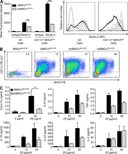
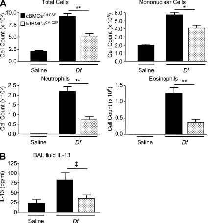
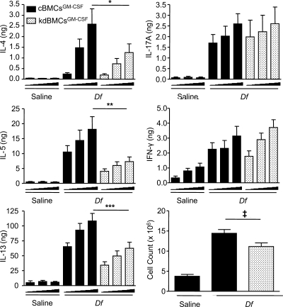
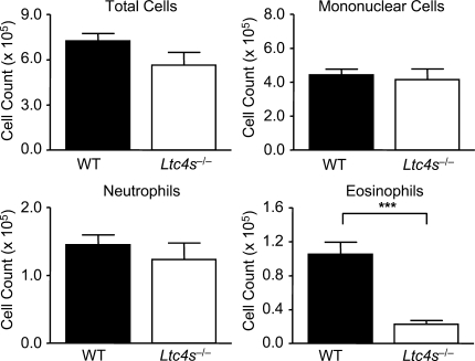
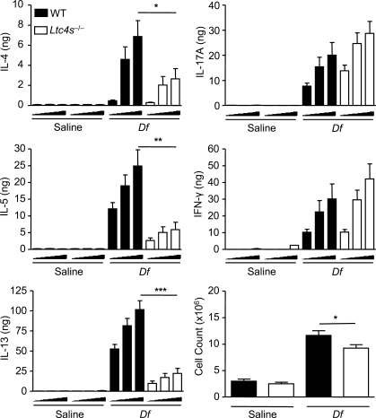
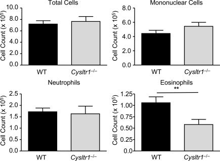
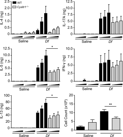
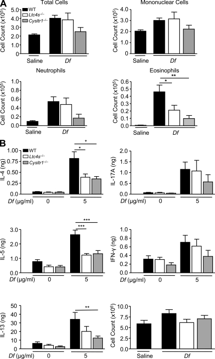
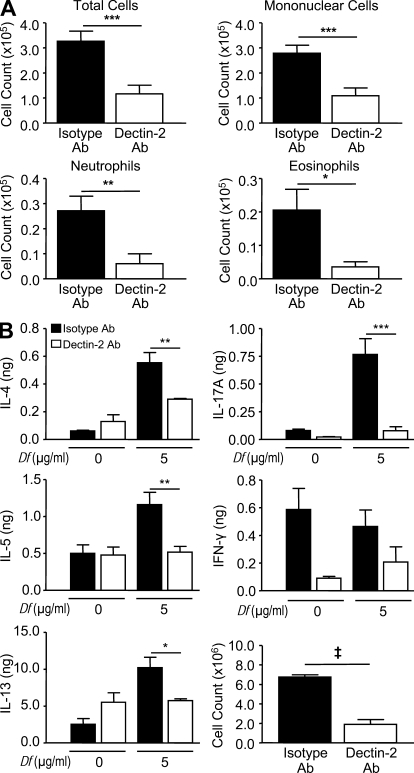
The innate signaling pathways for Th2 immunity activated by inhaled antigens are not well defined. We previously identified Dectin-2 as a receptor for glycans in allergen extracts from the house dust mite Dermatophagoides farinae (Df) that mediates cysteinyl leukotriene (cys-LT) generation from pulmonary CD11c+ cells and from GM-CSF-cultured bone marrow cells (BMCs(GM-CSF)). Using lentiviral knockdown of Dectin-2 in BMCs(GM-CSF) and adoptive transfer of Df-pulsed BMCs(GM-CSF) to sensitize naive mice, we now report that Dectin-2 is critical for the development of Df-elicited eosinophilic and neutrophilic pulmonary inflammation and Th2 cytokine generation in the lungs and restimulated lymph nodes. Sensitization with Df-pulsed BMCs(GM-CSF) from LTC(4) synthase (LTC(4)S)-deficient mice or type 1 cys-LT receptor (CysLT1R)-deficient mice demonstrated that both proteins were required for Df-elicited eosinophilic pulmonary inflammation and Th2 cytokine generation in the lungs and restimulated lymph nodes. Direct sensitization and challenge of Ltc4s-/- and Cysltr1-/- mice confirmed that cys-LTs mediate these parameters of Df-elicited Th2 pulmonary inflammation. Thus, the Dectin-2-cys-LT pathway is critical for the induction of Th2 immunity to a major allergen, in part through CysLT1R. These findings identify a previously unrecognized link between a myeloid C-type lectin receptor and Th2 immunity.


Figures

Figure 1.
Figure 1.
Dectin-2 mediates Df-elicited cytokine production by BMCsGM-CSF. BMCsGM-CSF were infected with viral particles containing Dectin-2 shRNA (kdBMCsGM-CSF), a vector control (cBMCsGM-CSF), or a nontargeting vector control (ntcBMCGM-CSF), selected with puromycin, and harvested at day 7. (A) Dectin-2 expression on all BMCsGM-CSF and on CD11chi MHCIIlow cells was assessed by flow cytometry. Left, results are means ± SEM (n = 4–6 mice per group) from three independent experiments. ***, P = 0.0001. Significance was determined with an unpaired Student’s t test. Right, a representative histogram of Dectin-2 expression on all BMCsGM-CSF and on CD11chiMHCIIlow cells from cBMCsGM-CSF (gray), ntcBMCGM-CSF (light gray), and kdBMCsGM-CSF (heavy black line) cultures is compared. Isotype control staining on all BMCsGM-CSF is also shown for cBMCsGM-CSF (gray dotted), ntcBMCsGM-CSF (light gray dotted), and kdBMCsGM-CSF (heavy black line dotted). (B) Representative histograms are shown of CD11c expression and MHCII expression on cBMCsGM-CSF, ntcBMCsGM-CSF, and kdBMCsGM-CSF, with the percentage of CD11c positive cells noted, as gated by the isotype control staining on a mixture of cBMCsGM-CSF, ntcBMCsGM-CSF, and kdBMCsGM-CSF, designated BMCsGM-CSF. MHCII staining was performed with nonsaturating antibody concentrations. Gating on a CD11chiMHCIIlow Dectin-2hi subset is also indicated (small boxes). (C) BMCsGM-CSF were stimulated with Df at the indicated concentrations and the supernatant contents were assessed by ELISA for cys-LTs at 60 min and for cytokines at 4 h. Results for cys-LT generation are means ± SEM (n = 6–8 mice per group) from four independent experiments. **, P = 0.02. Significance was determined with an unpaired Student’s t test. Results for cytokines are means ± SEM (n = 5–6 mice per group) from three independent experiments. P = 0.01 for IL-6, P = 0.04 for IL-10, P = 0.03 for IL-23, and P = 0.0001 for TNF. Significance was determined with two-way ANOVA (p-values refer to the integrated differences in cBMCsGM-CSF and kdBMCsGM-CSF over the varied doses).
Figure 2.
Figure 2.
BMCGM-CSF Dectin-2 is required for Df-elicited Th2 pulmonary inflammation. WT mice were sensitized by intranasal administration of 104 saline-pulsed or Df-pulsed kdBMCsGM-CSF or cBMCsGM-CSF at day 8 of culture, challenged with 2 µg Df on day 22 and day 23, and analyzed on day 25. (A) Cells from the BAL fluid were counted, cytospin preparations were stained, and 400 cells/slide were counted for specific cell types. Results are means ± SEM (n = 13–14 mice per group) from three independent experiments. **, P = 0.0001; *, P = 0.001. Significance was determined with an unpaired Student’s t test. (B) ELISA was performed on BAL fluid supernatants. Results are means ± SEM (n = 14 mice per group) from three independent experiments. ‡, P = 0.05.
Figure 3.
Figure 3.
BMCGM-CSF Dectin-2 is required for lymph node Th2 cytokine production elicited by Df. WT mice were sensitized with saline-pulsed or Df-pulsed kdBMCsGM-CSF or cBMCsGM-CSF and challenged with Df as described in the Fig. 2 legend. Parabronchial lymph node cells were isolated, counted, and restimulated for 72 h with 0, 1, or 5 µg/ml Df, and cytokines in the supernatant were measured by ELISA. Total cytokine production per mouse is shown. Results are means ± SEM (n = 10–13 mice per group) from three independent experiments. Triangles under x axes indicate 0, 1, and 5 µg/ml Df, from left to right. *, P = 0.04; **, P = 0.01; ***, P = 0.007; ‡, P = 0.03. Significance was determined with an unpaired Student’s t test.
Figure 4.
Figure 4.
BMCGM-CSF LTC4S is required for Df-elicited eosinophilic pulmonary inflammation. WT mice were sensitized by intranasal administration of 104 Df-pulsed WT or Ltc4s−/− BMCsGM-CSF, challenged with 2 µg Df on day 22 and day 23, and analyzed on day 25. Cells from the BAL fluid were counted, cytospin preparations were stained, and 400 cells/slide were counted for specific cell types. Results are means ± SEM (n = 13–14 mice per group) from four independent experiments. ***, P = 0.0001. Significance was determined with an unpaired Student’s t test.
Figure 5.
Figure 5.
BMCGM-CSF LTC4S is required for Df-elicited CD4+ Th2 cell recruitment to the lung. WT mice were sensitized with saline-pulsed or Df-pulsed WT or Ltc4s−/− BMCsGM-CSF and challenged with Df as described in the legend to Fig. 4. Pulmonary mononuclear cells were isolated, stimulated with 50 ng/ml PMA and 1 µM ionomycin for 10 h in the presence of 2.5 µM monensin, stained for cell surface expression of CD4 and CD8 (A) and for intracellular expression of IL-4, IL-5, IL-17A, and IFN-γ (B), and analyzed by flow cytometry. Gating on cell size, on CD4 and CD8 expression, the total number of cells recruited to the lung is shown. Results are means ± SEM (n = 11 mice per group) from two experiments. *, P = 0.02; **, P = 0.005; ***, P = 0.002; ****, P = 0.0008. Significance was determined with an unpaired Student’s t test.
Figure 6.
Figure 6.
BMCGM-CSF LTC4S is required for lymph node Th2 cytokine production elicited by Df. WT mice were sensitized with saline-pulsed or Df-pulsed WT or Ltc4s−/− BMCsGM-CSF and challenged with Df as described in the legend to Fig. 4. Parabronchial lymph node cells were isolated, counted, and restimulated for 72 h with 0, 1, or 5 µg/ml Df, and cytokines in the supernatant were measured by ELISA. Total cytokine production per mouse is shown. Results are means ± SEM (n = 12–15 mice per group) from three experiments. Triangles under x axes indicate 0, 1, and 5 µg/ml Df, from left to right. *, P = 0.04; **, P = 0.001; ***, P = 0.0001. Significance was determined with an unpaired Student’s t test.
Figure 7.
Figure 7.
Df-elicited eosinophilic pulmonary inflammation is dependent on BMCGM-CSF CysLT1R. WT mice were sensitized with intranasal administration of 104 Df-pulsed WT or Cysltr1−/− BMCsGM-CSF, challenged with 2 µg Df on day 22 and day 23, and analyzed on day 25. Cells from the BAL fluid were counted, cytospin preparations were stained, and 400 cells/slide were counted for specific cell types. Results are means ± SEM (n = 6–7 mice per group) from two experiments. **, P = 0.02. Significance was determined with an unpaired Student’s t test.
Figure 8.
Figure 8.
Df-elicited Th2 cytokine generation is dependent on BMCGM-CSF CysLT1R. WT mice were sensitized with saline-pulsed or Df-pulsed WT or Cysltr1−/− BMCsGM-CSF and challenged with Df as described in the legend to Fig. 7. Parabronchial lymph node cells were isolated, counted, and restimulated for 72 h with 0, 1, or 5 µg/ml Df, and cytokines in the supernatant were measured by ELISA. Total cytokine production per mouse is shown. Results are means ± SEM (n = 7 mice per group) from two experiments. Triangles under x axes indicate 0, 1, and 5 µg/ml Df, from left to right. *, P = 0.03; **, P = 0.02. Significance was determined with an unpaired Student’s t test.
Figure 9.
Figure 9.
Df-elicited eosinophilic pulmonary inflammation and Th2 cytokine generation is dependent on LTC4S and CysLT1R. WT (black), Ltc4s−/− (white), or Cysltr1−/− (gray) mice were sensitized with 10 µg Df or saline on days 0 and 1, challenged with 10 µg Df on days 15 and 16, and analyzed on day 18. (A) Cells from the BAL fluid were counted, cytospin preparations were stained, and 400 cells/slide were counted for specific cell types. Results are means ± SEM (n = 7–12 mice per group) from three experiments. *, P = 0.05; **, P = 0.01. (B) Parabronchial lymph node cells were isolated, counted, and restimulated for 72 h with 0 or 5 µg/ml Df, and cytokines in the supernatant were measured by ELISA. Total cytokine production per mouse is shown. Results are means ± SEM (n = 7–12 mice per group) from three experiments. *, P = 0.05; **, P = 0.01; ***, P = 0.001. Significance was determined with a one-way ANOVA.
Figure 10.
Figure 10.
Df-elicited pulmonary inflammation and Th2 cytokine generation is dependent on Dectin-2. WT mice were sensitized and challenged as described in the Fig. 9 legend. Mice were injected intraperitoneally with 200 µg Dectin-2 antibody or a rat IgG2a isotype control on days 0, 2, 15, and 17, and analyzed on day 18. (A) Cells from the BAL fluid were counted, cytospin preparations were stained, and 400 cells/slide were counted for specific cell types. (B) Parabronchial lymph node cell counts. Results are means ± SEM (n = 4 mice per group) and are representative of two independent experiments. *, P = 0.04; **, P = 0.02; ***, P = 0.01. (B) Parabronchial lymph node cells were isolated, counted, and restimulated for 72 h with 0 or 5 µg/ml Df, and cytokines in the supernatant were measured by ELISA. Total cytokine production per mouse is shown. Results are means ± SEM (n = 4 mice per group) and are representative of two independent experiments. *, P = 0.04; **, P = 0.03; ***, P = 0.01; ‡, P = 0.0001. Significance was determined with an unpaired Student’s t test.

References

    1. Altmann F. 2007. The role of protein glycosylation in allergy. Int. Arch. Allergy Immunol. 142:99–115 10.1159/000096114 - DOI - PubMed
    1. Aragane Y., Maeda A., Schwarz A., Tezuka T., Ariizumi K., Schwarz T. 2003. Involvement of dectin-2 in ultraviolet radiation-induced tolerance. J. Immunol. 171:3801–3807 - PubMed
    1. Ariizumi K., Shen G.L., Shikano S., Ritter R., III, Zukas P., Edelbaum D., Morita A., Takashima A. 2000. Cloning of a second dendritic cell-associated C-type lectin (dectin-2) and its alternatively spliced isoforms. J. Biol. Chem. 275:11957–11963 10.1074/jbc.275.16.11957 - DOI - PubMed
    1. Barrett N.A., Maekawa A., Rahman O.M., Austen K.F., Kanaoka Y. 2009. Dectin-2 recognition of house dust mite triggers cysteinyl leukotriene generation by dendritic cells. J. Immunol. 182:1119–1128 - PMC - PubMed
    1. Boehmler A.M., Drost A., Jaggy L., Seitz G., Wiesner T., Denzlinger C., Kanz L., Möhle R. 2009. The CysLT1 ligand leukotriene D4 supports α4β1- and α5β1-mediated adhesion and proliferation of CD34+ hematopoietic progenitor cells. J. Immunol. 182:6789–6798 10.4049/jimmunol.0801525 - DOI - PubMed

Publication types

MeSH terms