. 2011 Apr;60(4):1314-23.
doi: 10.2337/db10-1557. Epub 2011 Feb 28.

MicroRNA-200b regulates vascular endothelial growth factor-mediated alterations in diabetic retinopathy

Affiliations

MicroRNA-200b regulates vascular endothelial growth factor-mediated alterations in diabetic retinopathy

Kara McArthur et al. Diabetes. 2011 Apr.

Abstract

Objective: Diabetic retinopathy (DR) is a leading cause of blindness. Increased vascular endothelial growth factor (VEGF), promoting angiogenesis and increased permeability, is a key mechanistic abnormality in DR. We investigated microRNA (miRNA) alterations in DR with specific focus on miR-200b, and its downstream target, VEGF.

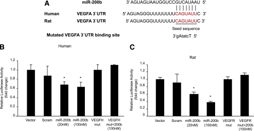
Research design and methods: miRNA expression profiling microarray was used to examine the retinas of streptozotocin-induced diabetic rats. Expressions of specific miRNAs were verified with PCR in the rat retina and in glucose-exposed endothelial cells. A target search, based on sequence complementarities, identified specific targets. We analyzed mRNA levels and protein expression in endothelial cells from large vessels and retinal capillaries and in the rat retina, with or without injection of miR-200b mimic or antagomir. Localization of miR-200b and its functional analysis in the rat and human retinas were performed.

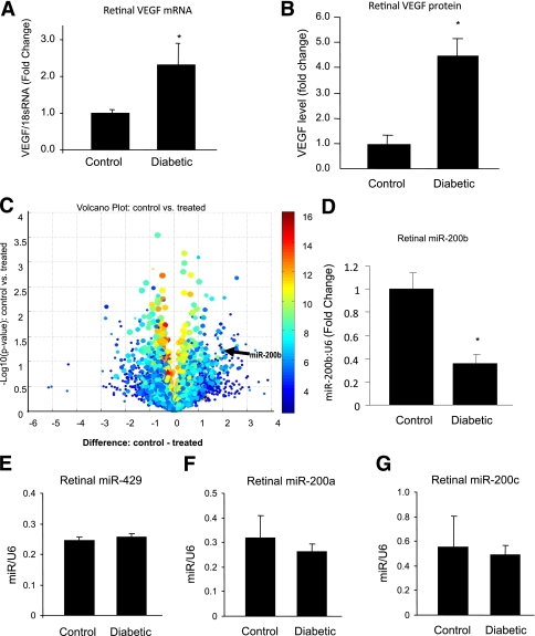
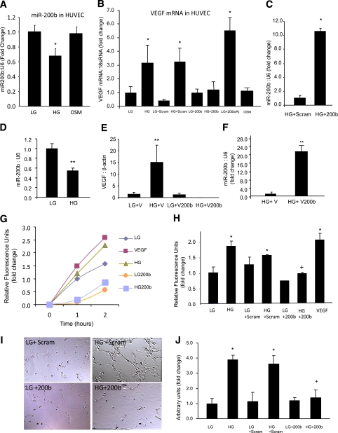
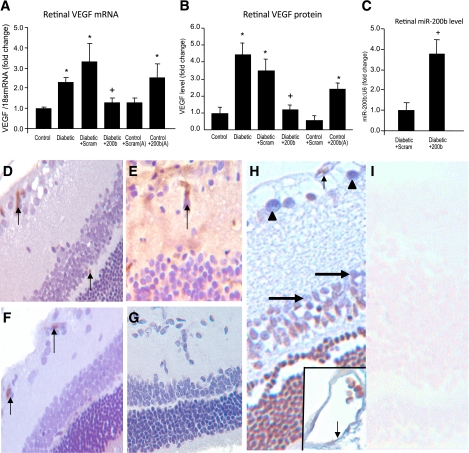
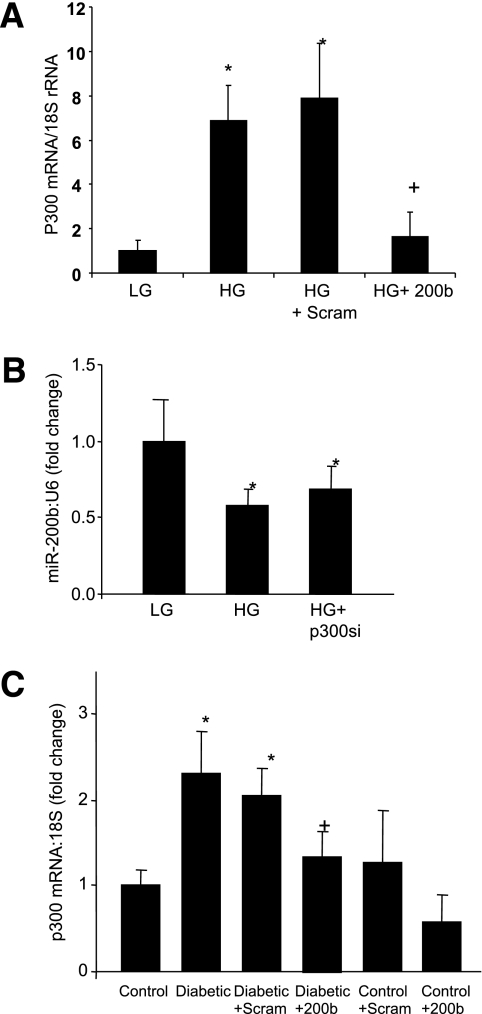
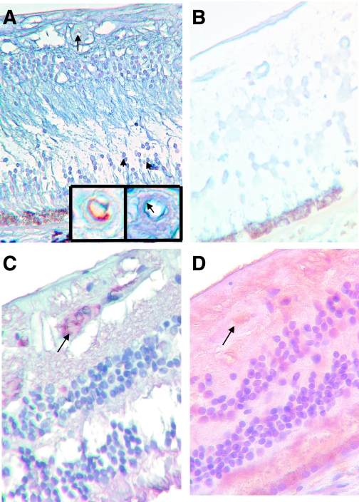
Results: Alteration of several miRNAs, including downregulation of miR-200b, were observed in the retina in diabetes. Such downregulation was validated in the retina of diabetic rats and in endothelial cells incubated in glucose. In parallel, VEGF (target of miR-200b) mRNA and protein were elevated. In the retina, miR-200b was localized in neuronal, glial, and vascular elements. Transfection of endothelial cells and intravitreal injection of miR-200b mimic prevented diabetes-induced increased VEGF mRNA and protein. Also prevented were glucose-induced increased permeability and angiogenesis. Furthermore, transfection of miR-200b antagonists (antagomir) led to increased VEGF production. Similar alterations were seen in the human retina.

Conclusions: These studies show a novel mechanism involving miR-200b in DR. Identification of such mechanisms may lead to the development of novel miRNA-based therapy.


Figures

FIG. 1.
FIG. 1.
MiRNA and VEGF alteration in the retina in diabetes. Quantitative RT-PCR (A) and ELISA (B) analysis from nondiabetic control and diabetic rat retinal tissue samples after 1 month of follow-up show increased levels of VEGF mRNA and protein. C: Volcano plot shows miRNA alteration in control vs. diabetic (treated) rat retina after 1 month. Circles to the left of −1 difference line and to the right of the 1 difference line are considered to have a fold-change >2× (x-axis is the log2 of the fold-change between two groups). The arrow indicates the location of miR-200b. (Custom analysis using Asuragen miRNA system. Some of the highly expressed miRNAs represent putative miRNA sites, which require biological characterization). Data array were validated with qRT-PCR analyses, which confirmed downregulation of miR-200b in the retina of diabetic rats compared with the nondiabetic controls (D) and no significant changes in the expression of miR-429 (E), miR-200a (F), and miR-200c (G). The miRNA data are expressed as a ratio to RNU6B (U6); mRNA levels are expressed as a ratio to 18S RNA, and normalized to controls. The error bars show the SEM. *Significantly different from the other group. (A high-quality color representation of this figure is available in the online issue.)
FIG. 2.
FIG. 2.
Effects of glucose-induced miR-200b downregulation in the endothelial cells. A: miR-200b was downregulated in the endothelial cells when exposed to 25 mmol/L d-glucose (HG) compared with 5 mmol/L d-glucose (LG), but not by 25 mmol/L l-glucose (osmotic control, OSM). B: Transfection of endothelial cells with miR-200b mimics (but not the scrambled mimics) normalized HG-induced upregulation of VEGF. Glucose-like effect (VEGF upregulation) was seen when cells in LG were transfected with miR-200b antagomir. OSM had no effect on VEGF expression. C: Efficiency of miR-200b mimic transfection as shown by increased miR-200b expression in the ECs after miR-200b mimic transfection compared with scrambled mimics. D: Similar to HUVECs, BRECs showed glucose-induced miR-200b downregulation. E: Transfection of BRECs with miR-200b mimics (using miR-200b cloned in PcDNA3.1 vector, but not by empty vector) normalized HG-induced upregulation of VEGF mRNA expression. F: Efficiency of miR-200b mimic transfection in the BREC was shown by increased miR-200b expression in these cells after miR-200b mimic transfection compared with vector control. HG-induced and VEGF-mediated increased transendothelial permeability (G), duration-dependent data and at the end point (H) were prevented by miR-200b mimic transfection. I: Similarly, glucose-induced EC tube formation was prevented by miR-200b mimic transfection. (Original magnification ×400.) J: The quantification of the tube formation assay is shown. Scram, scrambled miRNA; 200b, miR-200b mimic; 200b(A), 200b antagomir; V, PcDNA3.1 vecto. *Significantly different from LG or LG scram. +Significantly different from HG or HG scram. **Significantly different from other group(s). The miRNA levels are expressed as a ratio of RNU6B (U6) and normalized to LG; mRNA is expressed as a ratio to 18S RNA and normalized to LG. The error bars show the SEM. (A high-quality color representation of this figure is available in the online issue.)
FIG. 3.
FIG. 3.
A: Alignment of VEGF 3′-UTR (and mutated VEGF 3′-UTR) sequence with mature miR-200b based on bioinformatics predictions (www.TargetScan.org, www.microrna.org, www.ebi.ac.uk1). The 5′ end of the mature miR-200b is the seed sequence and has perfect complementarity with seven nucleotides of the 3′-UTR of VEGF. In the mutated sequence (small caps identifying mutated nucleotides) such complementarity was lost. B and C: Binding of miR-200b with VEGF promoter luciferase reporter assay shows dose-dependent binding of VEGF 3′-UTR with miR-200b, whereas mutated (mut) VEGF 3′-UTR abrogated the inhibitory effects of miR-200b. Relative promoter activities were expressed as luminescence units normalized for β-galactosidase expression. *Significantly different from vector, scrambled (scram) or mut. VEGFH, human VEGF; VEGFR, rat VEGF. (A high-quality color representation of this figure is available in the online issue.)
FIG. 4.
FIG. 4.
miR-200b–mediated alteration of retinal VEGF and its functional consequences. Levels of VEGF mRNA (A) and protein (B) in the control and diabetic rat retina, with or without intravitreal injection of miR-200b mimic (200b) and scrambled mimic (Scram) show that upregulated VEGF expression in the diabetic rat retina can be prevented by miR-200b mimics (but not by scrambled mimics). Intravitreal injection of miR-200b antagomir (200b[A]) produced VEGF upregulation. C: Efficiency of intravitreal delivery as demonstrated by increased retinal miR-200b expression after intravitreal injection of miR-200b mimic compared with scrambled mimic. *Significantly different from control. +Significantly different from diabetic or diabetic scram. The error bars show the SEM. D: Immunocytochemical stain on the control rat retina using antialbumin antibody shows only intravascular albumin (brown chromogen arrow; score = 0). E: Similar stain in the diabetic rat retina resulted in intravascular reactivity (arrow) and diffuse staining of the retina (score = 3), indicating increased vascular permeability. F: After intravitreal miR-200b injection, albumin staining was only present in the intravascular compartment (arrow; score = 0). G: Negative control of E shows specificity of staining. No such effects were seen after scrambled miR-200b injection (not shown). H: LNA-ISH study of retinal tissues in a control rat retina shows localization of miR-200b (blue chromogen) in the retinal capillaries (arrow), ganglion cells (arrowheads), and in the cells of inner nuclear layer (large arrow), both in the glial and neuronal elements. The inset shows an enlarged view of a capillary with miR-200b localization (arrow). I: LNA-ISH study of retinal tissues in a diabetic rat retina (in similar orientation) show minimum (if any) expression of miR-200b (ALK Phos was used as chromogen [blue] with no counterstain in LNA-ISH; DAB chromogen [brown] and hematoxylin counterstain in albumin stain). (Original magnification ×400 for DI.) (A high-quality digital representation of this figure is available in the online issue.)
FIG. 5.
FIG. 5.
miR-200b regulates diabetes-induced p300 alteration. A: miR-200b mimic (but not the scrambled mimics) transfection prevented glucose-induced p300 mRNA upregulation in the HUVECs (please see Fig. 2C regarding the efficiency of miR-200b mimic transfection). B: No effects of p300 siRNA transfection on miR-200b expression were seen. C: Augmented retinal p300 mRNA expression in the diabetic rats (compared with the controls) was prevented by intravitreal injection of miR-200b mimic (200b) but not by scrambled mimic (Scram). (Please see Fig. 4C regarding efficiency of intravitreal delivery). *Significantly different from control or LG (5 mmol/L d-glucose). +Significantly different from diabetic or HG (25 mmol/L d-glucose). The error bars show the SEM.
FIG. 6.
FIG. 6.
miR-200b alteration present in human diabetic retinopathy. A: LNA-ISH study of retinal tissues from nondiabetic human retina shows localization of miR-200b in the cells of the inner nuclear layer (arrowheads) and retinal capillaries (arrow). The inset (right) shows an enlarged view of the same capillary with endothelial localization of miR-200b (blue chromogen, arrow) and CD34 stain (brown chromogen) from an adjacent section showing endothelium of the capillary. B: Retinal tissues in a diabetic human retina (in similar orientation) show minimal (if any) expression of miR-200b. C: Immunocytochemical stain on the nondiabetic human retina using antialbumin antibody shows intravascular albumin (arrow). D: Diabetic human retina showed intravascular albumin staining (arrow) and diffuse staining of the retina, indicating increased vascular permeability. (ALK Phos was used as chromogen [blue] with no counterstain in LNA-ISH; DAB chromogen [brown] and hematoxylin counterstain in albumin stain.) (Original magnification ×400.) (A high-quality digital representation of this figure is available in the online issue.)

References

    1. Khan ZA, Farhangkhoee H, Chakrabarti S. Towards newer molecular targets for chronic diabetic complications. Curr Vasc Pharmacol 2006;4:45–57 - PubMed
    1. Brownlee M. Biochemistry and molecular cell biology of diabetic complications. Nature 2001;414:813–820 - PubMed
    1. Chuang JC, Jones PA. Epigenetics and microRNAs. Pediatr Res 2007;61:24R–29R - PubMed
    1. Latronico MV, Catalucci D, Condorelli G. MicroRNA and cardiac pathologies. Physiol Genomics 2008;34:239–242 - PubMed
    1. Liu J, Valencia-Sanchez MA, Hannon GJ, Parker R. MicroRNA-dependent localization of targeted mRNAs to mammalian P-bodies. Nat Cell Biol 2005;7:719–723 - PMC - PubMed

Publication types

MeSH terms