

Sexual dimorphism in the effect of concomitant progesterone administration on changes caused by long-term estrogen treatment in pituitary hormone immunoreactivities of rats
- PMID: 21358595
- PMCID: PMC3524720
- DOI: 10.12659/msm.881440
Sexual dimorphism in the effect of concomitant progesterone administration on changes caused by long-term estrogen treatment in pituitary hormone immunoreactivities of rats
Abstract
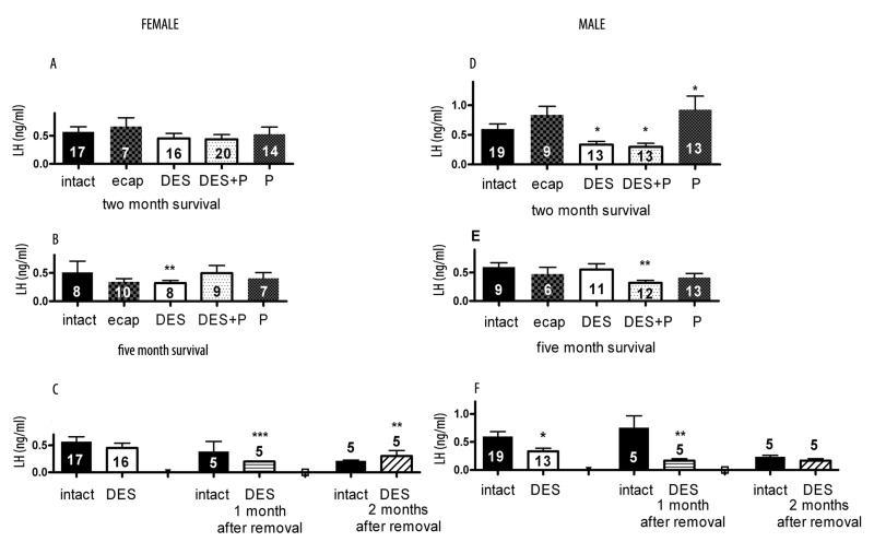
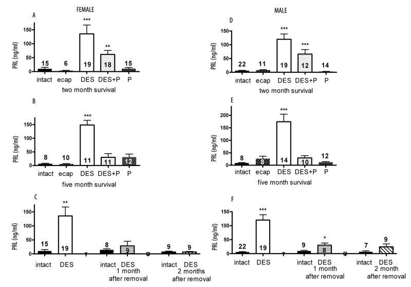
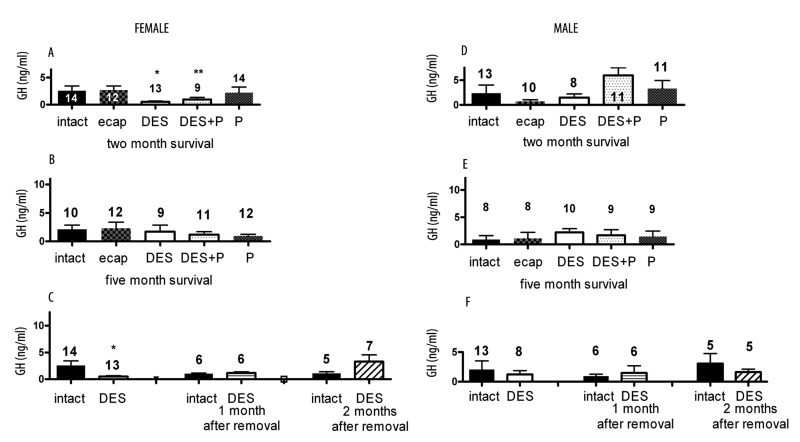
Background: Since in clinical practice long-term estrogen (E) treatment is frequently applied, our aim was to study the effect of concomitant progesterone (P) administration on changes caused by long-term estrogen treatment in the secretion of LH, FSH, PRL and GH.
Material/methods: Diethylstilbestrol (DES), P or both in silastic capsules were implanted under the skin of prepubertal Sprague-Dawley male and female rats. Animals survived for two or five months. We have also studied whether the changed hormone secretion caused by DES can return to normal level 1 or 2 months after removing DES capsule.
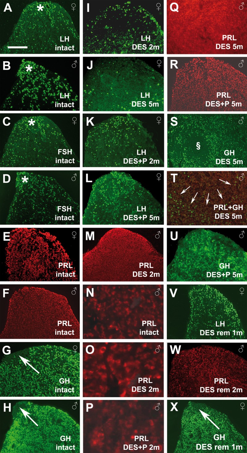
Results: 1.) The males more rapidly responded than females with decreasing basal LH release upon treatments. The basal FSH release was decreased only in males. The effect of DES persisted in males; however, in females basal LH and FSH levels were upregulated after removal of DES capsule. 2.) The basal GH levels were low in each group. The body weight and length were depressed by DES in both genders and P little blunted this effect. The body weight and length in males remained low after removal of DES capsule, in females it was nearly similar to intact rats. 3.) There was no sexual dimorphism in the effect of steroids on PRL secretion. In both genders DES extremely enhanced the PRL levels, P prevented the effect of DES. PRL levels returned to intact value after removal of DES influence. 4.) Removal of DES capsule reversed the changes in the immunohistochemical appearance of hormone immunoreactivities.
Conclusions: There was sexual dimorphism in the change of basal gonadotropic hormone and GH secretion but not of PRL upon DES and DES+P treatments. P was basically protective and this role may be mediated by P receptors locally in the pituitary gland.
Figures

Similar articles
-
Role of alpha-amino-3-hydroxy-5-methylisoxazole-4-propionic acid receptors in the control of prolactin, growth hormone and gonadotropin secretion in prepubertal rats.J Endocrinol. 1999 Sep;162(3):417-24. doi: 10.1677/joe.0.1620417. J Endocrinol. 1999. PMID: 10467233
-
Effect of concomitant progesterone administration or the effect of removal of estrogen capsule on changes caused by long-term estrogen treatment in pituitary VIP immunoreactivities.Endocrine. 2010 Jun;37(3):396-402. doi: 10.1007/s12020-010-9320-x. Epub 2010 Mar 30. Endocrine. 2010. PMID: 20960159
-
Effects of starvation in rats on serum levels of follicle stimulating hormone, luteinizing hormone, thyrotropin, growth hormone and prolactin; response to LH-releasing hormone and thyrotropin-releasing hormone.Endocrinology. 1977 Feb;100(2):580-7. doi: 10.1210/endo-100-2-580. Endocrinology. 1977. PMID: 401735
-
Neonatal exposure to DES in BALB/c male mice: effects on pituitary-gonadal function.Pharmacol Biochem Behav. 1985 Jun;22(6):1019-24. doi: 10.1016/0091-3057(85)90312-0. Pharmacol Biochem Behav. 1985. PMID: 3927323
-
Diethylstilbestrol increases the density of prolactin cells in male mouse pituitary by inducing proliferation of prolactin cells and transdifferentiation of gonadotropic cells.Histochem Cell Biol. 2006 Jul;126(1):111-23. doi: 10.1007/s00418-005-0141-6. Epub 2006 Feb 9. Histochem Cell Biol. 2006. PMID: 16468032
References
-
- Morgan WW, Steger RW, Smith MS, et al. Time course of induction of PRL-secreting pituitary tumors with diethylstilbestrol in male rats. Response of tuberoinfundibular dopaminergic neurons. Endocrinology. 1985;116:17–24. - PubMed
-
- Kuiper GGJM, Enmark E, Pelto-Huikko M, et al. Comparison of ligand binding specificity and transcript tissue distribution of estrogen receptors α and β. Proc Natl Acad Sc USA. 1996;93:5925–30. - PubMed
-
- Gonzales M, Reyes R, Damas C, et al. Oestrogen receptor α and β in female rat pituitary cells: An immunochemical study. Gen Comp Endocrinol. 2008;155:857–68. - PubMed
-
- Wilson ME, Price RH, Jr, Handa RJ. Estrogen receptor beta messenger ribonucleic acid expression in the pituitary gland. Endocrinology. 1998;139:5151–56. - PubMed
Publication types
MeSH terms
Substances
LinkOut - more resources
Full Text Sources
