

Relation between expression pattern of p53 and survivin in cutaneous basal cell carcinomas
- PMID: 21358596
- PMCID: PMC3524735
- DOI: 10.12659/msm.881442
Relation between expression pattern of p53 and survivin in cutaneous basal cell carcinomas
Abstract
Background: The tumor suppressor gene p53 is a key regulator of cell division and/or apoptosis. Survivin is a multifunctional member of the inhibitor of apoptosis family. Survivin and p53 represent diametrically opposed signals that influence the apoptotic pathway.
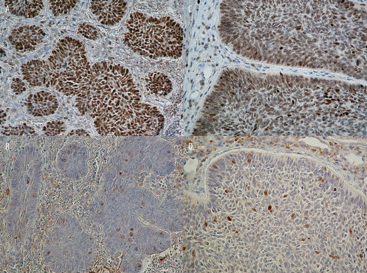
Material/methods: To determine the role of p53 and survivin in basal cell carcinoma (BCC), we evaluated the expression pattern of both proteins with regard to the percentage of positively immunostained tumor cells, the intensity of staining, and subcellular localization among 31 subjects with BCC.
Results: Overexpression of p53 protein was found in 28 of 31 cases (90.3%), whereas survivin accumulation was seen in 27 (87.1%). For p53, moderate and/or strong immunoreactivity was seen in 20 of 28 cases (71.4%), and 26 of 28 cases (92.9%) showed more than 25% reactive tumor cells. Nuclear p53 staining was detected in 23 of 28 cases (82.1%), whereas combined nuclear and cytoplasmic localization was found in only 5 of 28 cases (17.9%). Survivin revealed mild intensity of immunoreaction in 22 of 27 cases (71%), and 25 of 27 cases (92.6%) showed less than 25% labeled tumor cells. Combined nuclear and cytoplasmic survivin localization was present in 26 of 27 cases (96.3%). Statistically significant differences were detected in the assessed expression parameters between those proteins.
Conclusions: Our results suggest that overexpression of wild type p53 protein may suppress the expression of survivin and its antiapoptotic activity in BCC cells.
Figures
References
-
- Kastan MB, Onkyekwere O, Sidransky D, et al. Participation of p53 protein in the cellular response to DNA damage. Cancer Res. 1991;51:6304–11. - PubMed
-
- Mirza A, McGuirk M, Hockenberry TN, et al. Human survivin is negatively regulated by wild-type p53 and participates in p53-dependent apoptotic pathway. Oncogene. 2002;21:2613–22. - PubMed
-
- Hoffman WH, Biade S, Zilfou JT, et al. Transcriptional Repression of the Anti-apoptotic survivin Gene by Wild Type p53. J Biol Chem. 2002;277:3247–57. - PubMed
-
- Momand J, Wu HH, Dasgupta G. MDM2-master regulator of the p53 tumor supressor protein. Gene. 2000;242:15–29. - PubMed
-
- Fritsche M, Haessler C, Brandner G. Induction of nuclear accumulation of the tumor-supressor protein p53 by DNA-damaging agents. Oncogene. 1993;8:307–18. - PubMed
Publication types
MeSH terms
Substances
LinkOut - more resources
Full Text Sources
Medical
Research Materials
Miscellaneous
