. 2011 Mar;29(3):267-72.
doi: 10.1038/nbt.1788. Epub 2011 Feb 27.

Generation of anterior foregut endoderm from human embryonic and induced pluripotent stem cells

Affiliations

Generation of anterior foregut endoderm from human embryonic and induced pluripotent stem cells

Michael D Green et al. Nat Biotechnol. 2011 Mar.

Abstract

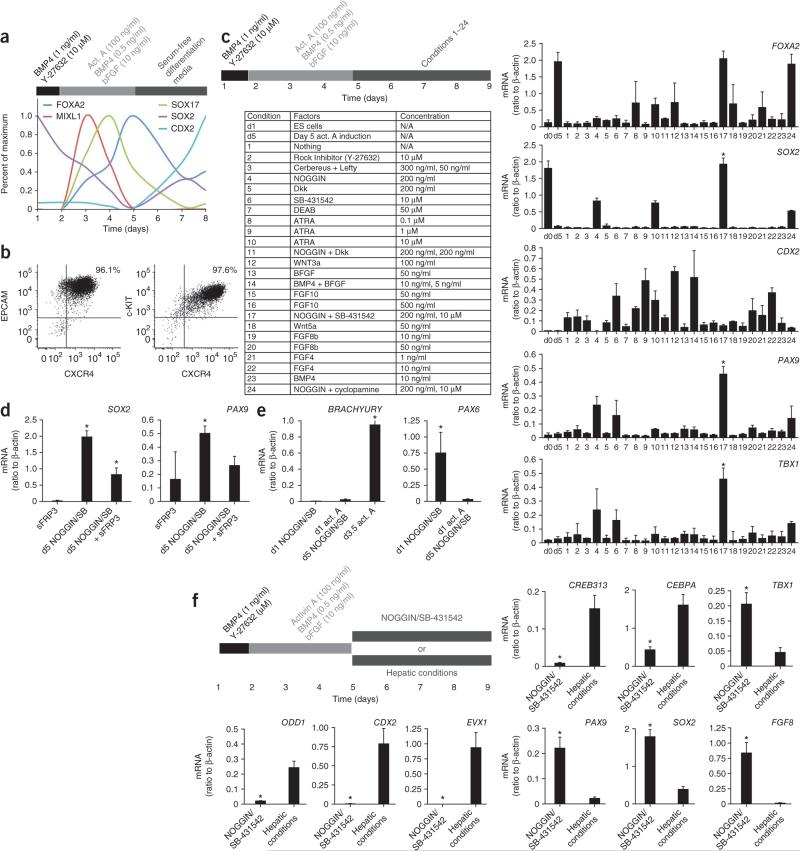
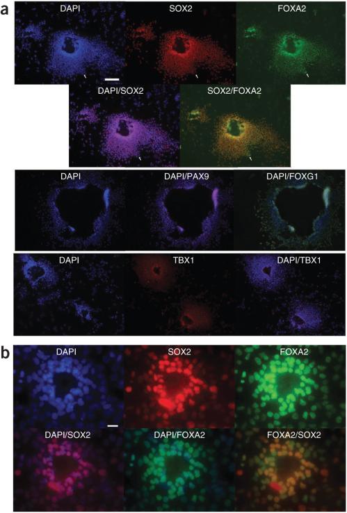
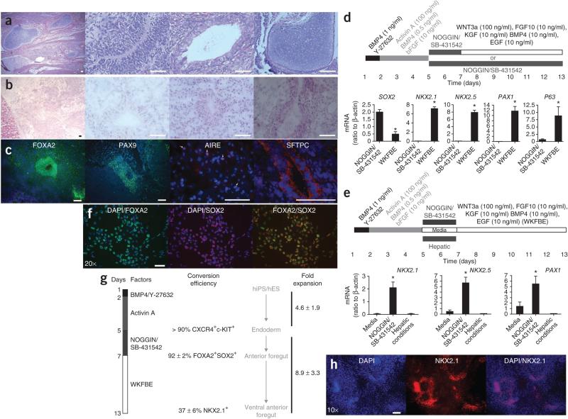
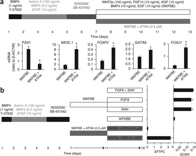
Directed differentiation of human embryonic stem (hES) cells and human induced pluripotent stem (hiPS) cells captures in vivo developmental pathways for specifying lineages in vitro, thus avoiding perturbation of the genome with exogenous genetic material. Thus far, derivation of endodermal lineages has focused predominantly on hepatocytes, pancreatic endocrine cells and intestinal cells. The ability to differentiate pluripotent cells into anterior foregut endoderm (AFE) derivatives would expand their utility for cell therapy and basic research to tissues important for immune function, such as the thymus; for metabolism, such as thyroid and parathyroid; and for respiratory function, such as trachea and lung. We find that dual inhibition of transforming growth factor (TGF)-β and bone morphogenic protein (BMP) signaling after specification of definitive endoderm from pluripotent cells results in a highly enriched AFE population that is competent to be patterned along dorsoventral and anteroposterior axes. These findings provide an approach for the generation of AFE derivatives.


Figures

Figure 1
Figure 1
Induction of AFE markers in NOGGIN/SB-431542-treated definitive endoderm. (a) Expression of FOXA2, MIXL1, SOX17, SOX2 and CDX2 mRNA during activin A–mediated induction of definitive endoderm in hES cells. Data expressed as quantification of mRNA normalized to β-ACTIN (also known as ACTB), scaled proportionally to maximum induction. Cytokines were added as indicated on top of the figure (bar). (b) Representative flow cytometric analysis of definitive endodermal markers CXCR4, C-KIT and EPCAM at day 5 of activin A induction. Two biologically independent experiments are shown. (c) Expression of FOXA2, SOX2, CDX2, PAX9 and TBX1 mRNA on day 9 in cultures treated on day 5 after induction of definitive endoderm (see upper left panel), with the factors listed in the lower left panel (n = 3 biological replicates; *, significantly different from all other conditions, P < 0.0001; one-way ANOVA). d0, prior to start of differentiation; d5, day 5. (d) Expression of SOX2 and PAX9 on day 9 in cultures treated on day 5, after induction of definitive endoderm, with NOGGIN/SB-431542 (SB) in the presence or absence of sFRP3 (*, P < 0.05, n = 3 biological replicates). (e) Expression of BRACHYURY and PAX6 mRNA at day 9 in hES cells differentiated as previously described to neurectoderm (day 1 addition of NOGGIN/SB-431542), or after induction of endoderm (endoderm induction until day 5, followed by addition of NOGGIN/SB-431542). For BRAYCHURY, day 3.5 hES cells exposed to activin A and undergoing gastrulation served as a positive control (*, P < 0.0001, n = 3 experiments consisting each of three biological replicates). (f) Expression of ODD1, CDX2, EVX1, CREB313, CEBPA, TBX1, PAX9, SOX2 and FGF8 mRNA in day 9 cultures treated in parallel with either NOGGIN/SB-431542 or cultured in hepatic conditions after induction of definitive endoderm until day 5 (n = 3 experiments consisting each of three biological replicates).
Figure 2
Figure 2
Immunofluorescence analysis of NOGGIN/SB-431542-treated definitive endoderm. (a) Immunofluorescence for FOXA2, SOX2, CDX2, PAX9, FOXG1 and TBX1 of day 9 in HES2 definitive endoderm cultures treated on day 5 with NOGGIN/SB-431542. Scale bar, 50 μm (upper); 100 μm (lower). (b) Expression of FOXA2 and SOX2 in HFD9 hiPS cultures in the same conditions. Scale bar, 25 μm.
Figure 3
Figure 3
Functional characteristics of NOGGIN/SB-431542-induced AFE cells. (a) H/E staining of a teratoma derived after 5 weeks from HES2 transplanted under the kidney capsule of NOD/SCIDIl2rg−/− mice. The three right-hand panels show neurectoderm (neural rosette), endoderm (intestinal epithelium) and mesoderm (cartilage), respectively. Scale bar, 50 μ. (b) H/E staining of a growth arising 5 weeks after transplantation of NOGGIN/SB-431542-induced AFE cells derived from HES2 cells under the kidney capsule of immunocompromised mice. Scale bar, 50 μm. (c) Immunofluorescence analysis of the tissue from b stained for FOXA2, PAX9, AIRE and SFTPC. Scale bar, 50 μm. (d) Expression of SOX2, NKX2.1, NKX2.5, PAX1 and P63 in HES2-derived cells generated in the two conditions schematically represented on top of the panel (n = 6 culture wells from two independent experiments; *, significantly different from NOGGIN/SB-431542; P < 0.05) WKFBE: WNT3a, KGF, FGF10, BMP4 and EGF. (e) Expression of NKX2.1, NKX2.5 and PAX1 in HES2-derived cells generated in the three conditions schematically represented on top of the panel (n = 4 to 6 culture wells from three independent experiments, *, significantly different from the other conditions; P < 0.05). (f) Expression of FOXA2 (green) and SOX2 (red) 2 d after treatment of activin A–induced definitive endoderm with NOGGIN/SB-431542 (blue, DAPI). Scale bar, 50 μm. (g) Schematic overview of the efficiency of induction of ventral AFE. WKFBE: WNT3a, KGF, FGF10, BMP4 and EGF. (h) Immunofluorescence for NKX2.1 in differentiated HDF9 hiPS cells after sequential treatment with activin A, NOGGIN/SB-431542 and WKFBE according to the scheme in g. Scale bar, 50 μm.
Figure 4
Figure 4
Induction of lung and pharyngeal pouch markers from ventral AFE generated in vitro. (a) Expression of PAX1, NKX2.1, FOXP2, GATA6 and FOXJ1 in HES2-derived cells generated in the two conditions schematically represented on top of the panel (n = 4 to six culture wells from three independent experiments, *, significantly different from WKFBE conditions; P < 0.05). WKFBE: WNT3a, KGF, FGF10, BMP4 and EGF. (b) Induction of SFTPC and GCM2 mRNA in ventralized AFE in the presence of factors indicated in the figure.

References

    1. D'Amour KA, et al. Production of pancreatic hormone-expressing endocrine cells from human embryonic stem cells. Nat. Biotechnol. 2006;24:1392–1401. - PubMed
    1. Gouon-Evans V, et al. BMP-4 is required for hepatic specification of mouse embryonic stem cell-derived definitive endoderm. Nat. Biotechnol. 2006;24:1402–1411. - PubMed
    1. Cai J, et al. Directed differentiation of human embryonic stem cells into functional hepatic cells. Hepatology. 2007;45:1229–1239. - PubMed
    1. Murry CE, Keller G. Differentiation of embryonic stem cells to clinically relevant populations: lessons from embryonic development. Cell. 2008;132:661–680. - PubMed
    1. Spence JR, et al. Directed differentiation of human pluripotent stem cells into intestinal tissue in vitro. Nature. 2010;470:105–109. - PMC - PubMed

Publication types